
|
|
Benign nodular hyperplasia - Case 21.
|
|


First examination (first row of images)
Clinical data: a 56-year-old man was referred for an evaluation of a recurrent nodular goiter. He was operated 14 years ago, histopathology resulted in benign, hyperplastic nodules. He had no complaints.
Palpation: a multinodular goiter.
Functional state: euthyroidism (TSH 0.69 mIU/L, FT4 12.4 pM/L).
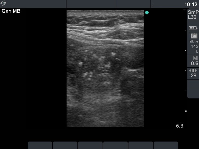
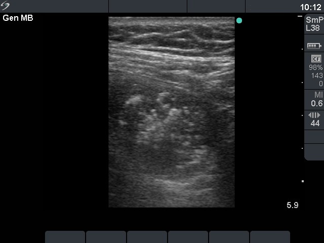
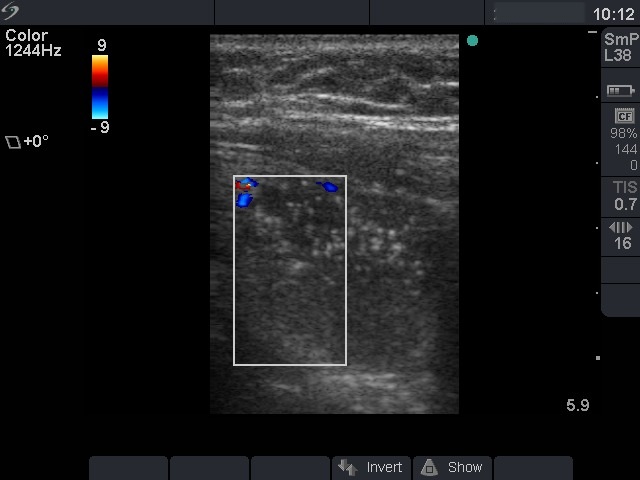
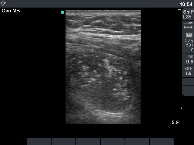
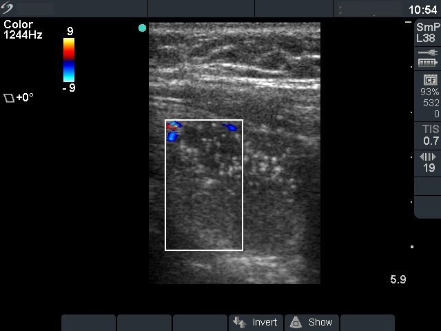
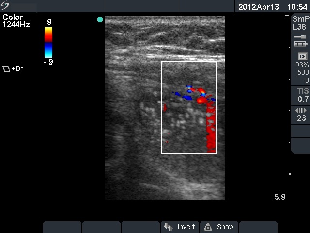
Ultrasonography: both thyroids were enlarged and echonormal. There were multiple, moderately hypoechogenic nodules in both lobes. There was a hypoechogenic nodule in the central part of the left lobe which presented hyperechogenic patches containing bright, hyperechogenic punctate granules. This pattern is similar to that observed in medullary cancer. The vascularization was not specific.
FNAC: was repeatedly not diagnostic.
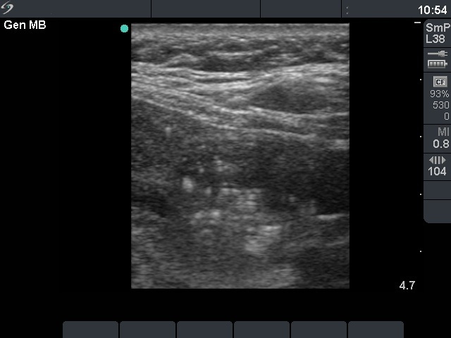
Second examination 14 months later (second row of images)
The whole thyroid increased in volume by 38%. Surgery was advised
Histopathology: benign hyperplastic nodular goiter.