Benign nodular hyperplasia - Case 58. (ultrasonographic picture 1)

|
|
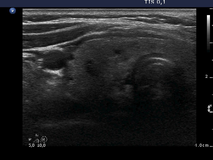
Right lobe, horizontal scan. There are several small cystic lesions within echonormal background.

|
|
|
Right lobe, horizontal scan. There are several small cystic lesions within echonormal background.