
|
|
The role of complex diagnosis - follow-up of follicular lesions - Case 9.
|
|

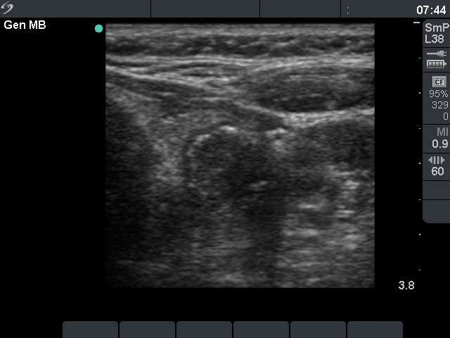
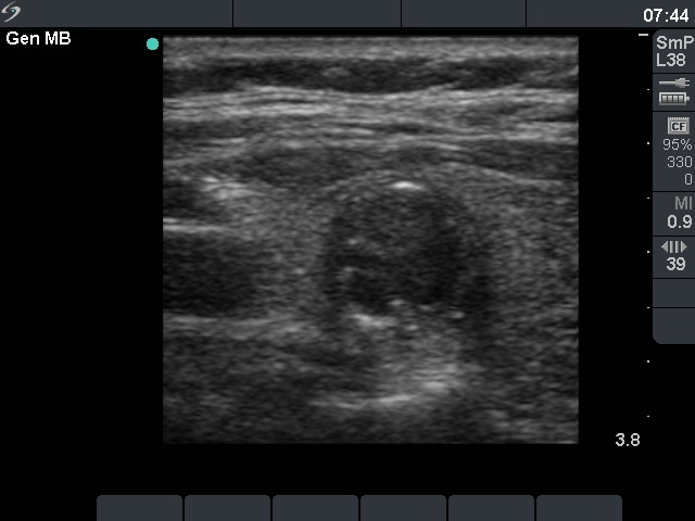


First examination (1st and 2nd rows of images)
Clinical presentation: A 69-year-old woman was referred for evaluation of nodular goiter. She was treated for kidney insufficiency. The nodule was discovered on regular checking.
Palpation: no abnormality.
Functional state: euthyroidism with TSH-level 0.56 mIU/L.
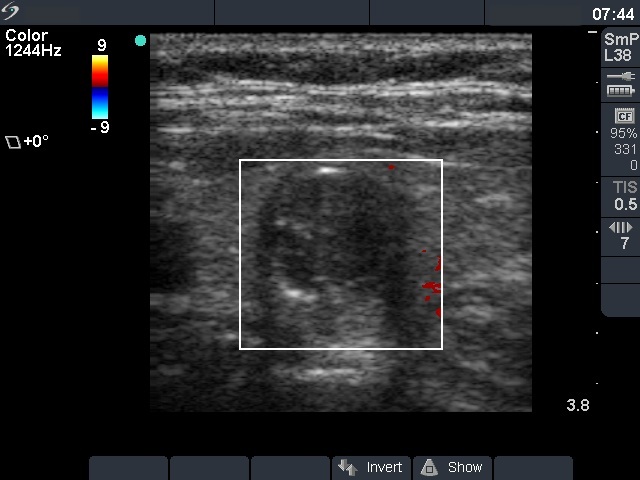
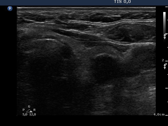
Ultrasonography. The thyroid was echonormal. There was a minimally hypoechogenic nodule in the right and a hypoechogenic nodule in the left lobe. The latter presented the so-called eggshell calcification.
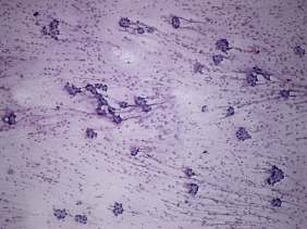
Cytology was performed and resulted in follicular tumor without significant atypia.
A combined clinical-ultrasound-cytological diagnosis was follicular tumor with less than 1% risk of carcinoma.
We advised instead of surgery regular follow-up examinations.
Follow-up examination 4 years later (3rd row of images)
Summary of follow-up: the patient underwent yearly ultrasound examination. The nodule was unchanged, she had no complaints.
Functional state: euthyroidism with TSH-level 1.94 mIU/L.
Ultrasonography: The ultrasound presentation of the thyroid was unchanged.
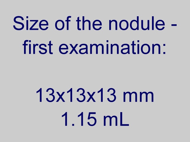
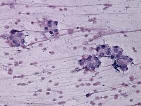
The volume of the nodule was 1.15 mL and 1.43 mL, at the first examination and at the 4-year follow up, respectively. It means a 24% increase in volume which is beyond the intraobserver variation.Aspiration cytology was repeated and resulted in benign follicular proliferation.
Suggestion: continuation of the follow-up with ultrasound and TSH determinations every year.