
Benign nodular hyperplasia - Case 25. (ultrasonographic picture 3)
|
|

|
|
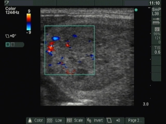
Right lobe, longitudinal scan, color Doppler method. Type 2 vascular pattern, i.e. increased perinodular blood flow. The intranodular vascularization is not specific.