
Papillary carcinoma - Case 1. (ultrasound picture 5)
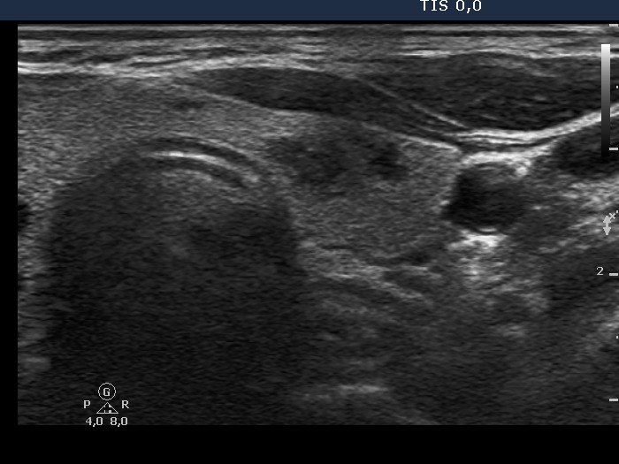 Lower part of the left lobe, horizontal view. A hypoechogenic, inhomogeneous lesion is demonstrated in this image. This nodule displays blurred borders. |
Papillary carcinoma - Case 1. (ultrasound picture 5)
Lower part of the left lobe, horizontal view. A hypoechogenic, inhomogeneous lesion is demonstrated in this image. This nodule displays blurred borders. |


