
|
|
Ethanol sclerotherapy: other examples - Case 3: treatment of a gelatinous thyroid cyst
|
|


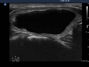


Three month prior to sclerotherapy (1st row of images)
Clinical presentation: a 50-year-old woman noticed a lump in the neck for 3 months. Thesize of the lesion did not changed over time.
Palpation: a firm nodule in the left lobe. The lesion was painless.
Functional state: euthyroidism with TSH 1.08 mIU/L.
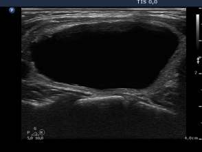
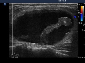
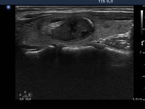
Ultrasonography: the thyroids were echonormal. A large central-type cyst occupied almost the entire left lobe.
We tried to remove the cystic fluid but only 1.5 ml brown gelatinous fluid could be aspirated. Aspiration cytology resulted in benign cystic lesion.
We suggested a repeat aspiration 3 months later.
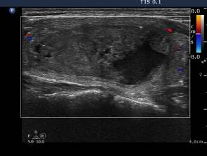
First session of sclerotherapy (second row of images):
Clinical presentation: the patient was treated by another endocrinologist after surgery. The patient was well and euthyroid on daily 87.5 microgram levo-tiroxine replacement therapy. She noticed a lump in her right thyroid for 3 months. She was advised to undergo repeated surgical procedure.
Palpation: unchanged.
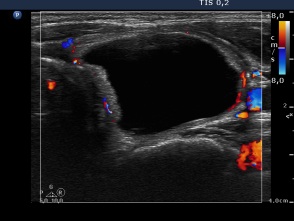
Ultrasonography remained unchanged.
We could aspirate 2 ml thick, brown cystic fluid and injected 3 mL ethanol.
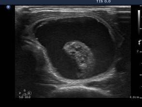
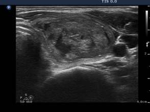
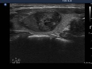
Second session of sclerotherapy (third row of images):
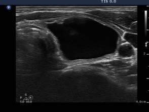
The second session was performed nine days after the previous one. The nodule became a bit larger. Now we could aspirated 15 mL brown fluid form the nodule, therafter we injected 5 mL ethanol.
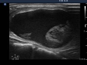
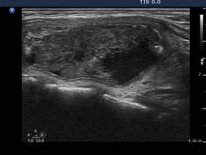
Third session of sclerotherapy (fourth row of images):
The lesion decreased in size. After removal of 2 mL bloody fluid we injected 4 mL ethanol.
Follow-up investigations 6 weeks after the last session (fifth row of images):
The lesion significantly decreased in size. We planned 2 more sessions but the patient asked me whether further sessions are absolutely necessary or not. I explained her that further sessions increase the long-term success of the therapy but we can postpone the decision considering the follow-up results. She decided not to be treated at this time.
Follow-up investigations 6 months after the last session (sixth row of images):
Clinical presentation: the patient did not feel the nodule. Her difficulties in swallowing have ceased.
Ultrasonography: a further significant decrease was detected. The nodule became more inhomogeneous.
Suggestion: repeat ultasound in one year.
Comment. Around one-tenth of larger non-aspirable cyst become aspirable spontaneously over time while this figure is even greater (exceeding 50%) if we administer ethanol after an unsuccessful attermpt of removal of cystic fluid.