
Benign nodular hyperplasia - Case 27. (ultrasonographic picture 1)

|
|
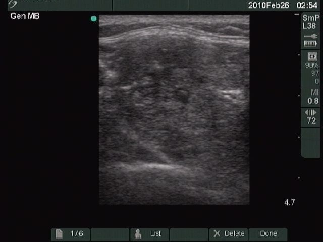
Right lobe, horizontal scan. The nodule occupying almost the entire lobe has a mixed structure: partly moderately hypoechogenic, partly hyperechogenic. This is a typical benign pattern.