
Benign nodular hyperplasia - Case 17. (ultrasonographic picture 2)

|

|

|
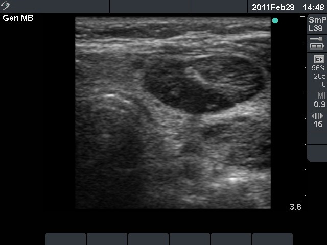
Left lobe, horizontal scan. A hypoechogenic nodule in the ventral and a moderately hypoechogenic nodule in the dorsolateral part. The acoustic shadow best seen on the dorsal part of the former lesion discloses the presence of coarse calcification.


