Benign nodular hyperplasia - Case 17. (ultrasonographic picture 4)

|

|

|
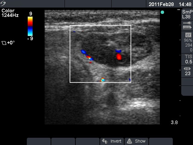
Left lobe, horizontal scan, color Doppler method. The largest nodule displays signs of type 3 vascular pattern.

|
|
|
|
Left lobe, horizontal scan, color Doppler method. The largest nodule displays signs of type 3 vascular pattern.


