
|
|
Other rare tumors and non-thyroidal lesions in the region of the thyroid - Case 13.Hyalinizing trabecular adenoma |
|


Clinical presentation: A 48-year-old man was referred for an evaluation of a nodular goiter which has been known for at least 4 years. The thyroid gradually increased in size. The patient had had difficulties in swallowing for 6 months.
Palpation: A firm nodule occupied almost the entire right thyroid.
Laboratory tests: euthyroidism with TSH 0.52 mIU/L, FT4 15.8 pM/L.
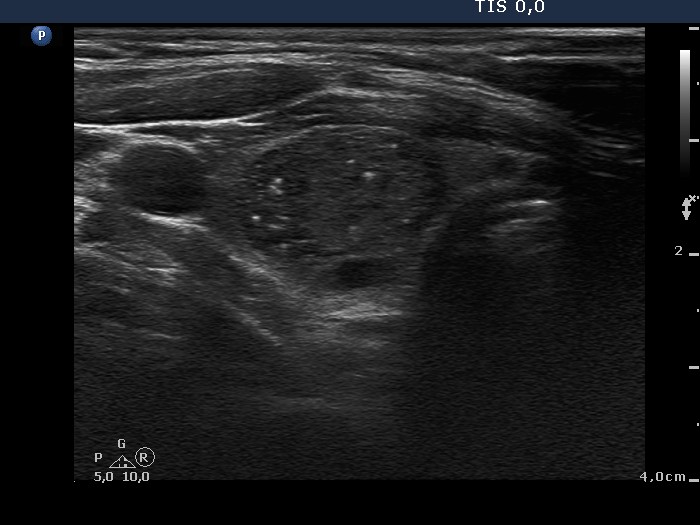
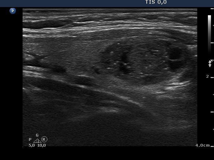
Ultrasonography: There were multiple hypoechogenic and moderately hypoechogenic nodules in both lobes. They contained small hyperechogenic granules, more probably comet-tail artifacts than microcalcifications. The largest lesion in the lower part of the right lobe was aspirated. The nodule displayed a combined perinodular and intranodular vascular pattern.
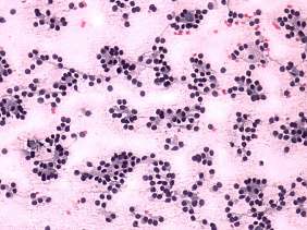
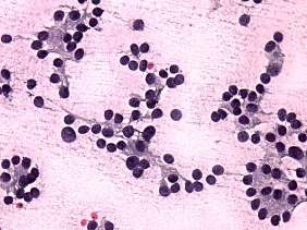
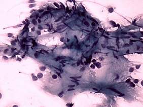
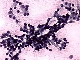
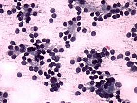
Cytological diagnosis: follicular tumor.
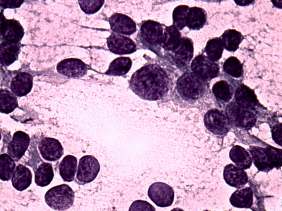
Histopathology: hyalinizing trabecular adenoma of the thyroid.
Comments: It is worth analyzing the cellular arrangement. Thyrocytes occur in three different structures: in follicles, in trabecular groups and in syncytial clusters with hyaline stroma.