
|
|
Papillary carcinoma - Case 70.
|
|

Clinical data: a 73-year-old woman was requested an evaluation of suspected hyperthyroidism.
Palpation: a hard nodule was palpable in the right side of the isthmus.
Functional state: euthyroidism (TSH 0.61 mIU/L, FT4 21.1 pM/L).
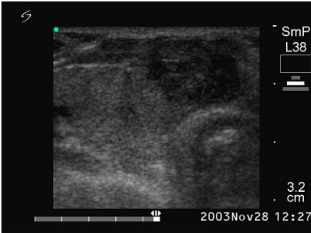
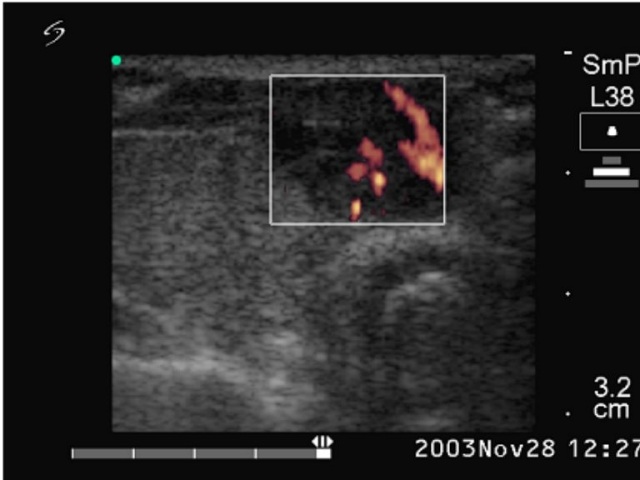
Ultrasonography: echonormal thyroids with two nodules close to each other in the isthmus. They were hypoechogenic inhomogeneous and exhibited irregularly increased intranodular blood flow.
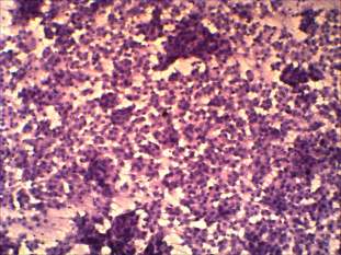
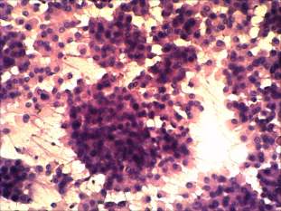
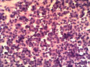
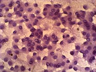
Cytological picture: no colloid in the background. Extremely cellular picture. Thyrocytes in irregular clusters, in microfollicles and predominantly dissociated. Almost all cells exhibit oxyphilic metaplasia and prominent nucleoli. Many cells contain groove. The size of the cells is relatively uniform.
Cytological diagnosis: Hürthle-cell tumor.
Histopathology: Hürthle-cell variant of papillary carcinoma.