
Papillary carcinoma - Case 29. (ultrasonographic picture 2)

|

|

|
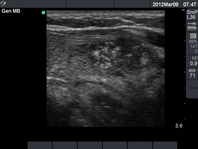
Left lobe, longitudinal view. A hypoechogenic lesion with patchy hyperechogenic areas and microcalcifications. The borders of the nodule are irregular.


