
|
|
Papillary carcinoma - Case 29.
|

|


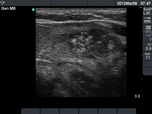


Clinical presentation: a 58-year-old woman was referred for an evaluation of a nodular goiter noticed for several months.
Palpation: a firm nodule in the left lobe.
Functional state: euthyroidism (TSH-level 3.01 mIU/L).
Ultrasonography: the thyroids were minimally-moderately hypoechogenic. The left lobe contained several echonormal lesions without any oncological significance. There was a large nodule in the lower part of the left thyroid. It was composed of multiple lesions. One of the latter was hypoechogenic, inhomogeneous and presented microcalcifications. Both the perinodular and the intranodular blood flow was increased.
Cytological diagnosis: suspicion of a Hürthle-cell tumor.
Histopathology: chronic lymphocytic thyroiditis. Benign hyperplastic nodules in the left lobe and a papillary cancer with a maximal diameter of 13 mm.
Comments:
-
It is worth thoroughly analyzing the cytological images. To raise the possibility of a carcinoma because of cytological reasons was a very hard task. The most important sign is the irregular arrangement of thyrocytes. There are neither grooves nor inclusions present. Nevertheless, the cytological pattern itself is not fully enough to raise the possibility of malignancy.
-
I am in a more convenient situation because I myself perform ultrasonography. To analyze a smear with limited signs of malignancy facing with this sonographic pattern was not as hard as would be without having an ultrasound presentation.


