
|
|
Papillary carcinoma - Case 30.
|

|


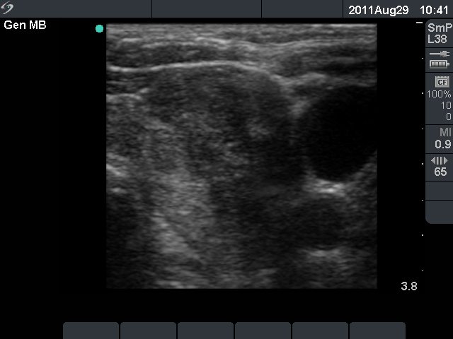

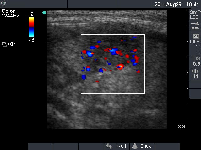




Clinical presentation: a 77-year-old woman was referred for an evaluation of a nodular goiter known for decades. The patient had no compression signs.
Palpation: a multinodular goiter.
Functional state: euthyroidism (TSH-level 0.94 mIU/L, FT4 12.6 pM/L).
Ultrasonography: an enlarged multinodular goiter was seen. There was a hypoechogenic nodule in the ventrolateral part of the left lobe. It had irregular borders and presented increased intranodular blood flow. There were multiple lymph nodes in the left side of the neck.
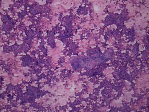
Cytological diagnosis: papillary cancer - both from the hypoechogenic lesion and from the lymph node.
Histopathology: disclosed papillary carcinoma in the left lobe with metastasis to the regional lymph node s.
Comment: the identical twin-sister of the patient was evaluated 2 years later. There is a startling resemblance between these two cases, including not only the final diagnosis, but also the presentation and location of the nodules. See case 4 of papillary carcinomas.


