
|
|
Papillary carcinoma - Case 31.
|
|


Clinical presentation: a 49-year-old woman was referred for an evaluation of suspected hypothyroidism.
Palpation: both thyroids were firm and a firmer nodule was palpable in the left lobe.
Functional state: subclinical hypothyroidism (TSH-level 8.76 mIU/L, FT4 12.2 pM/L).
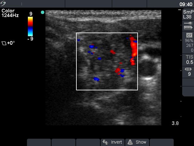
Ultrasonography: the thyroids were hypoechogenic and inhomogeneous. There was a more inhomogeneous nodule in the upper part of left lobe. The nodule contained microcalcifications and presented intranodular blood flow.
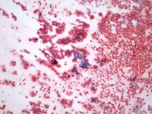
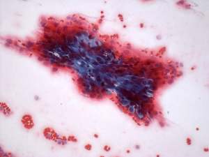
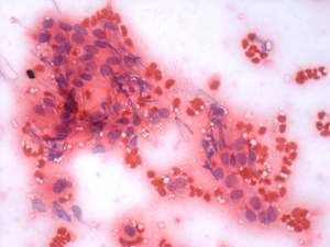
Cytological diagnosis: suspicion of papillary cancer.
Combined diagnosis: subclinical hypothyroidism caused by Hashimoto's thyroiditis. Suspicion of papillary cancer in the left lobe.
Histopathology: Hashimoto's thyroiditis and a papillary cancer in the left lobe.
Comment: it is worth comparing the insignificant discrete lesions detected in the right thyroid and the nodule in the left lobe on the video. Note the difference in echogenicity and vascularization.