
|
|
Chronic lymphocytic thyroiditis - Case 56.
|
|


Clinical presentation: a 40-year-old woman was referred for an evaluation of a thyroid nodule that had been discovered on carotid Doppler examination.
Palpation: both thyroids were firm, but no nodule was palpable.
Hormonal investigation: indicated euthyroidism with TSH-level 3.52 mIU/L.
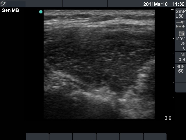
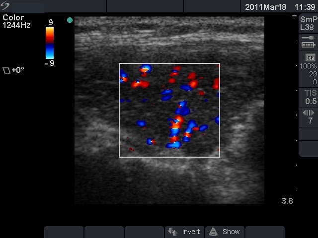
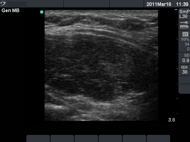
Ultrasonography: revealed hypoechogenic inhomogeneous thyroids with an echonormal circumscribed lesion in the left lobe corresponding to a secondary lobule.
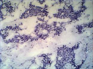
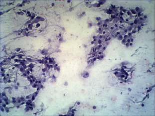
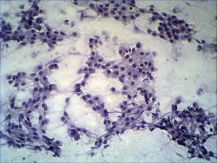
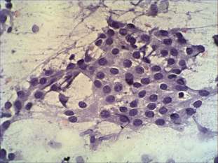
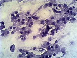
Cytological picture: oxyphilic cells predominate the smear. They had prominent nucleoli, several atypical enlarged cells were also present. The very small number of dissociated cells, the presence of nuclear debris and lymphocytes were against the possibility of tumor, but neoplasm could not be excluded solely on the cytological picture.
Combined ultrasonographic-cytological diagnosis: benign Hashimoto's thyroiditis.
Follow-up investigations: one year later subclinical hypothyroidism developed. The sonographic picture had also changed: the discrete lesion in the left lobe disappeared.