
Benign nodular hyperplasia - Case 50. (ultrasonographic picture 4)

|

|

|
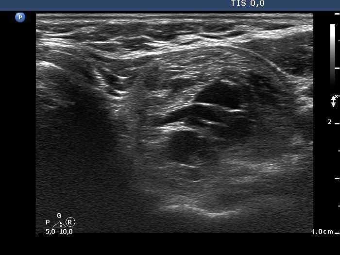
Upper part of the left lobe, horizontal scan. There is a mixed, hyperechogenic-cystic nodule in the central part of the lobe. The lesion has typical presentations of posterior back wall enhancement.


