
Benign nodular hyperplasia - Case 50. (ultrasonographic picture 5)

|

|

|
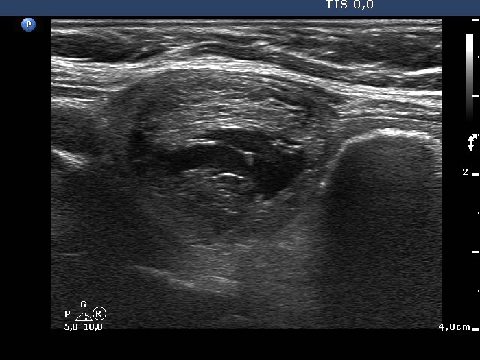
Upper part of the left lobe, longitudinal scan. The hyperechogneic lines ventral to the cystic area are fibrotic changes while the hyperechogenic figures dorsal to the cystic area are the consequence of posterior back wall enhancement.


