
Benign nodular hyperplasia - Case 51. (ultrasonographic picture 5)
|
|

|
|
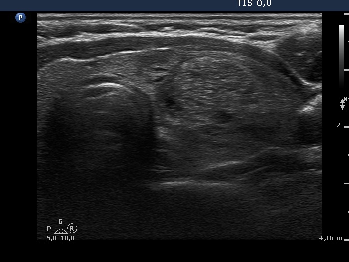
Left lobe, another horizontal scan. There is a relatively large echonormal nodule presenting fibrosis and non-specific hyperechogenic figures.