Benign nodular hyperplasia - Case 7. (ultrasonographic picture 5)

|

|

|
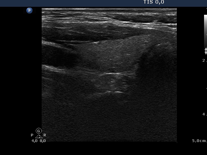
Left lobe, longitudinal scan. The volume of the left lobe became less than 1/4 of the average.

|
|
|
|
Left lobe, longitudinal scan. The volume of the left lobe became less than 1/4 of the average.


