
|
|
Papillary carcinoma - Case 6.
|
|


Clinical data: a 33-year-old woman was referred for an evaluation of a nodule discovered by herself several weeks earlier.
Palpation: a hard nodule in the left lobe.
Functional state: euthyroidism (TSH-level 4.60 mIU/L).
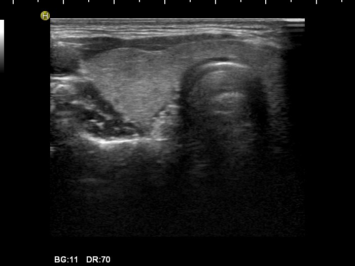
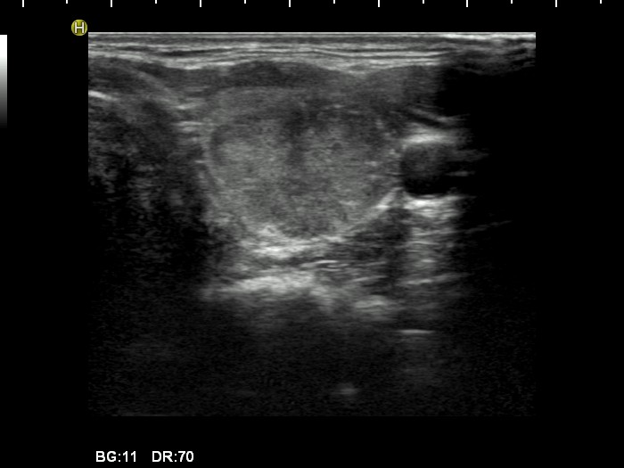
Ultrasonography: the thyroids were echonormal. There was a hypoechogenic nodule in the ventral part of the left lobe. The borders of the nodule were irregular, the lesion contained microcalcifications and displayed an irregularly increased type 3 vascular pattern.
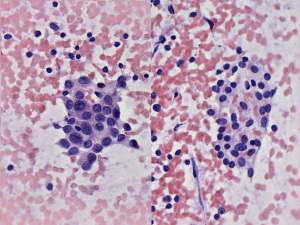
Cytological diagnosis: papillary cancer. Lymphocytic thyroiditis.
Histopathology: papillary cancer with focal lymphocytic thyroiditis. Metastases to the submandibular lymph nodes.
Comments:
-
The sonographic pattern is highly suspicious for papillary cancer. All of the important sonographic properties specific for this type of cancer are presented: hypoechogenicity, irregular borders, microcalcifications, an irregular type 3 vascular pattern.
-
It is worth analyzing the cytological images - two cell populations were found. Firstly, normal thyroid follicular cells, secondly follicular cells displaying the features of papillary cancer.