
Benign nodular hyperplasia - Case 19. (ultrasonographic picture 2)
|
|

|
|
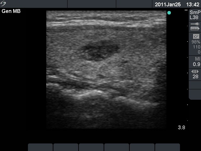
Right lobe, longitudinal scan. A hypoechogenic nodule with hyperechogenic granules. The latter are not as bright as in the case of a microcalcification.