
Benign nodular hyperplasia - Case 42. (ultrasonographic picture 1)

|
|
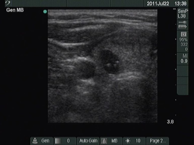
Right lobe, horizontal scan. A hypoechogenic nodule with hyperechogenic granules which have a dorsal tail; therefore these figures are colloid crystals.