
Papillary carcinoma - Case 68. (ultrasonographic picture 1)

|
|
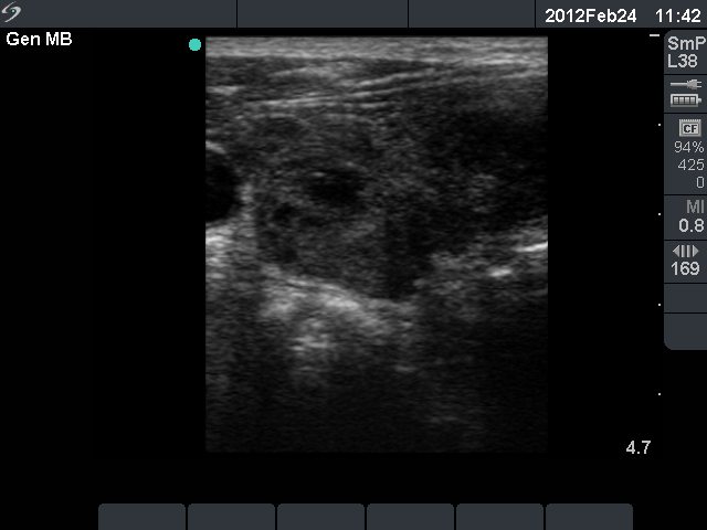
Right lobe, horizontal scan. The right thyroid is presented on the left side of the image: an echonormal thyroid with around 50% of hypoechogenic areas - this is one of most typical sonographic presentation of Hashimoto's thyroiditis.
The large hypoechogenic lesion in the isthmus can be identified on the right side of the picture.