
|
|
Chronic lymphocytic thyroiditis - Case 51.
|
|


Clinical presentation: a 38-year-old woman requested an evaluation in another thyroid outpatient department of complaints of hyperthyroidism. A hypoechogenic suspicious nodule was detected on ultrasonography. FNAC resulted in oxyphilic variant of papillary cancer. The patient requested a second opinion.
Results of blood tests: subclinical hypothyroidism with TSH-level 8.04 mIU/L, FT4 11.7 pM/L.
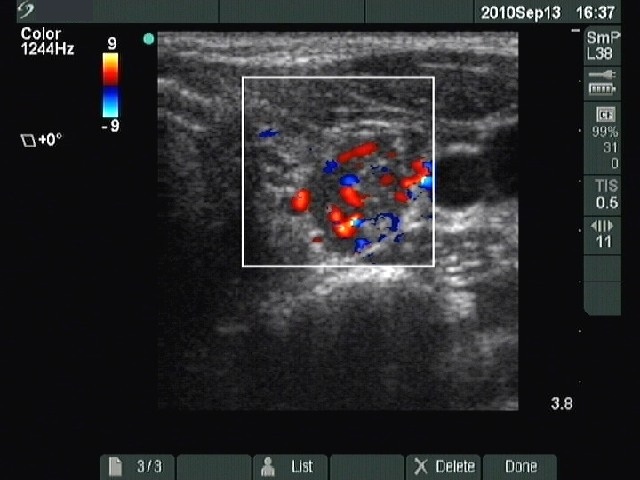
Ultrasonography: the basic echo structure of the thyroid was echonormal. There were hypoechogenic nodule-like lesions in both lobes.
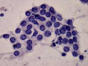
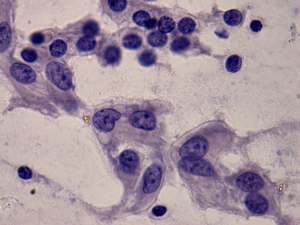
Cytological picture: there is no colloid in the background. Clusters of follicular cells. Thyrocytes show oxyphilic metaplasia, vary in size, but no significant atypia is present, they do not present prominent nucleoli. Clusters composed of oxyphilic cells mixed with lymphocytes were also found. The proportion of lymphocytes was relatively low. Clusters of elongated fibrotic elements was also present on the smear.
Cytological diagnosis: benign, Hashimoto's thyroiditis.
Although we suggested follow-up investigation instead of surgery, we could not resolve the anxiety caused by the result of the first aspiration cytology. Therefore, we agreed with the patient to undergo on surgery.
Histopathology: Hashimoto's thyroiditis without any nodule.