Benign cystic nodule (cytology and wash-out) - case 2175
|
Transverse scan |
Longitudinal scan |
 |
 |
 |
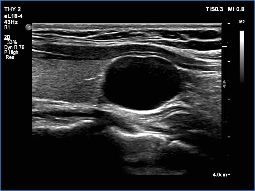 |
This lesion is almost completely cystic nodule. It is not evident whether the solid-looking areas (arrows) at the inner wall of the cyst correspond indeed to solid thyroid tissue or are clots.
Since the lesion has completely disappeared after aspiration, this should be clearly considered a purely cyst.
|
| |
|
|
Transverse scan |
Longitudinal scan |
 |
 |
 |
 |
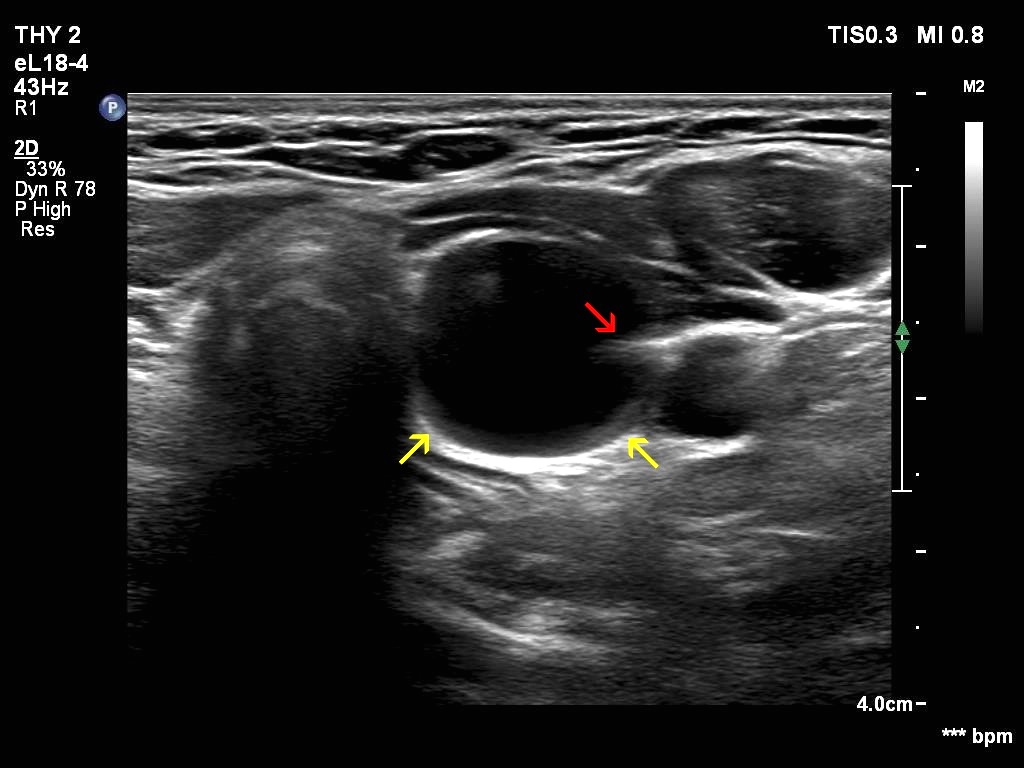
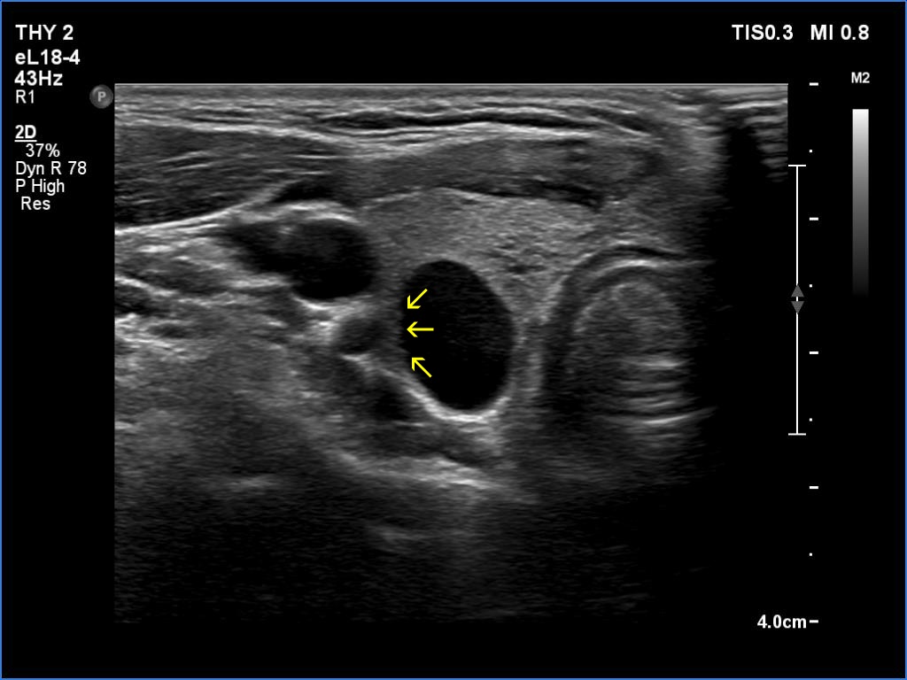
This is an almost completely cystic lesion, however it does not meet the criteria of a pure cyst. The wall thickening (yellow arrows) is ambiguous but there is a tiny solid-appearing area in the dorsal wall (red arrows). In systems which do not use the term 'almost completely cystic lesion', this nodule should be categorized as a peripheral-type cyst.
|
| |
|
| |
|
Benign cystic nodule (cytology and wash-out) - case 2177
|
Transverse scan |
Longitudinal scan |
 |
 |
 |
 |
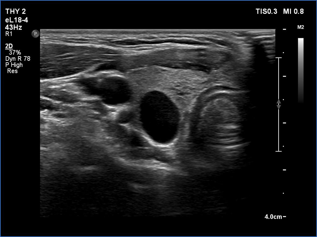
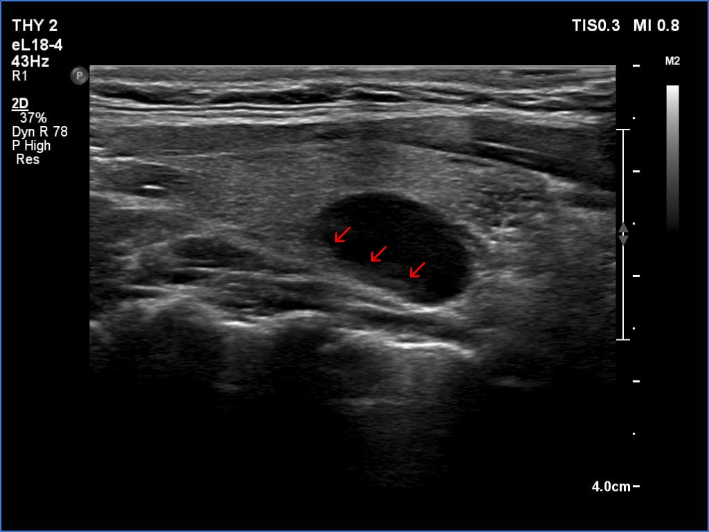
According to the EU-TIRADS, this lesion cannot be regarded as a purely cystic nodule due to the presence of wall-thickening (yellow arrows). The EU-TIRADS defines this lesion as an EU-TIRADS 3 nodule.
|
| |
|
|
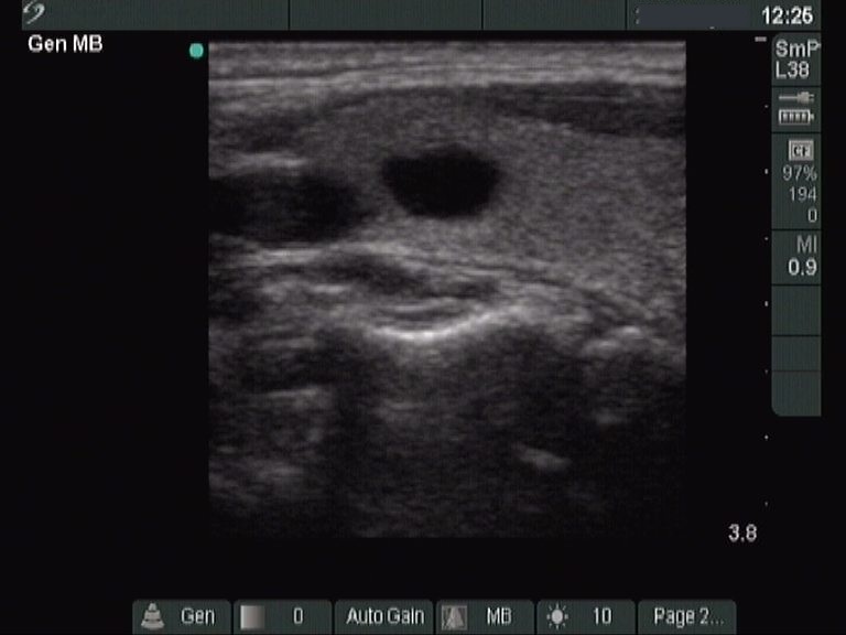
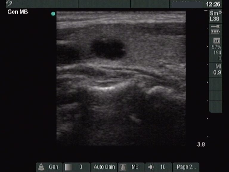
Before the aspiration |
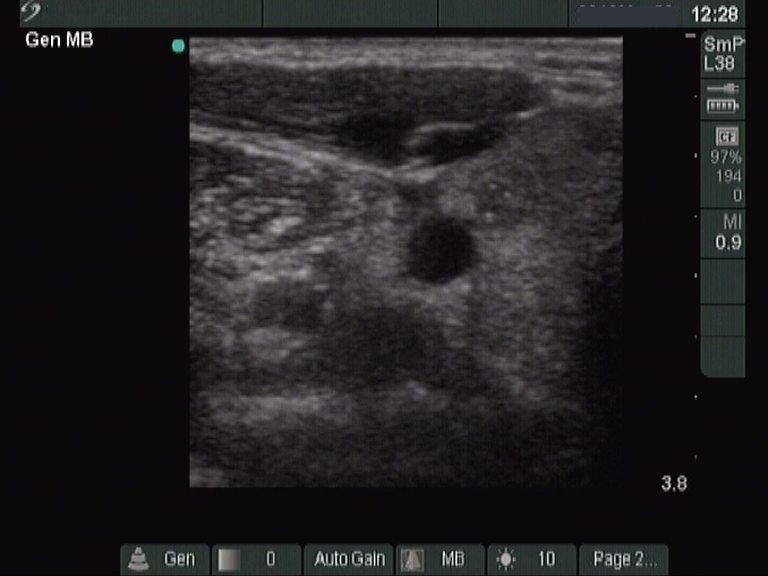
After the aspiration |
 |
 |
 |
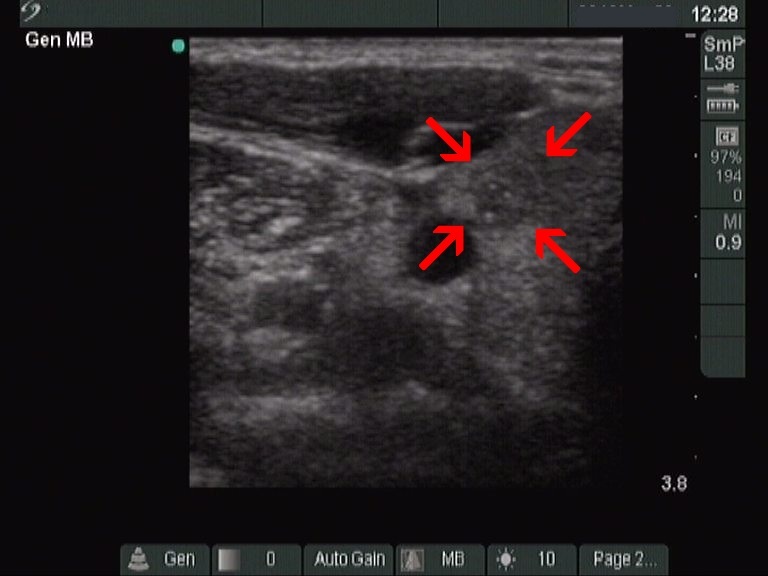 |
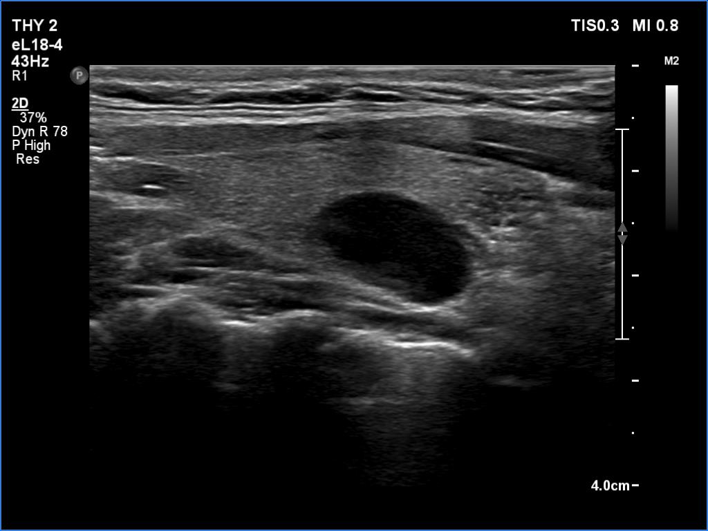
This case draws attention to why aspiration may also make sense in seemingly pure cysts, moreover it advocates the strict definition of pure cyst of the EU-ITRADS. The solid area containing microcalcifications (marked with red arrows) became visible only after the aspiration of fluid. Nevertheless, a very thin solid part could be found at the ventral wall even before the aspiration.
|
| |
|
Benign cystic nodule (ultrasound diagnosis) - case 28
|
Transverse scan |
Longitudinal scan |
 |
 |
 |
 |
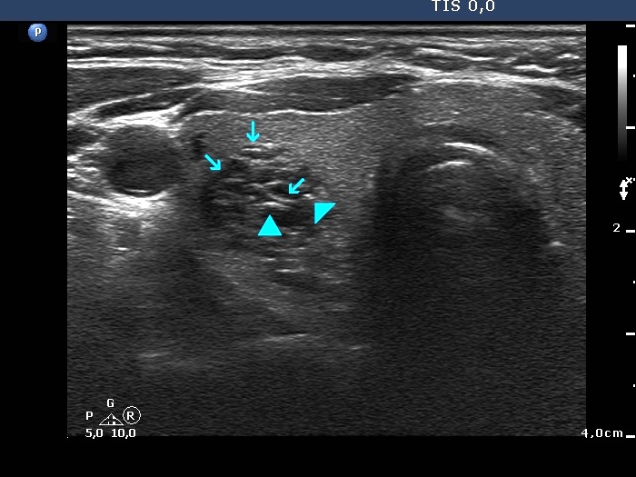
This lesion is very close to the spongiform sphere defined by EU_TIRADS. Nevertheless, the non-cystic part of the nodule involves not only fibrous septa but also a minimal solid portion, as well. Note the posterior acoustic enhancement in the dorsal wall of the small cystic areas. These causes no concern if this optical artifact is linear (arrows), however granular figures might be misinterpreted as microcalcifications (arrowhead).
|
| |
|
|
 |
 |
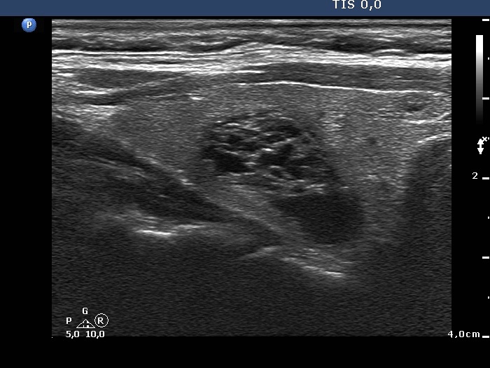
The extent of spongiform areas exceeds 50%, but not the entire nodule is spongiform. Therefore, it depends on the definition whether this nodule should be held as spongiform or not.
Since, according to the ETA, only a nodule with fully spongiform parts can be classified as a spongiform lesion, this cannot be considered as such.
|
| |
|
| |
|
| |
|
| |
|
