|
|
 |
 |
 |
 |
|
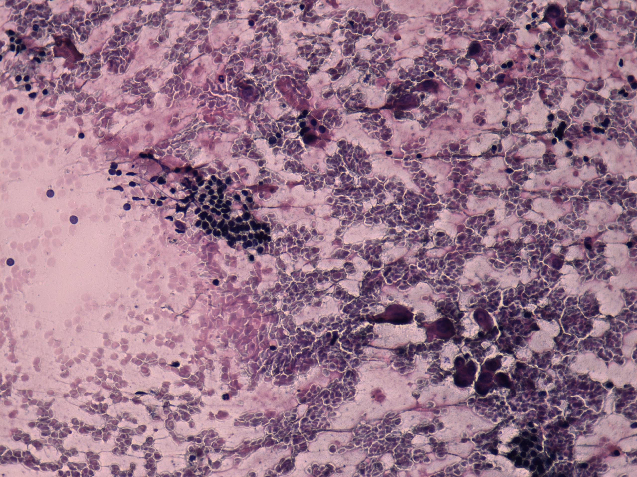
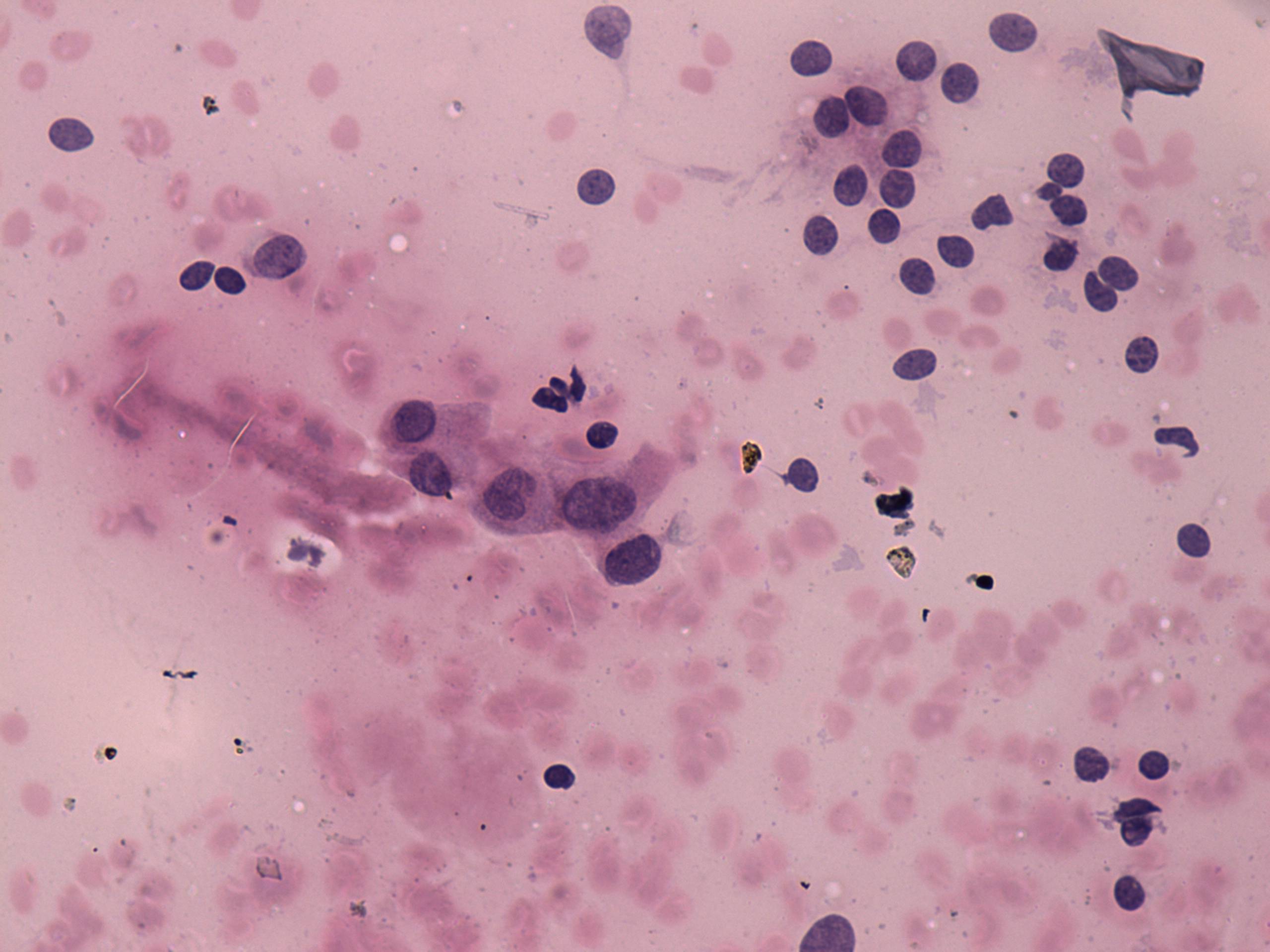
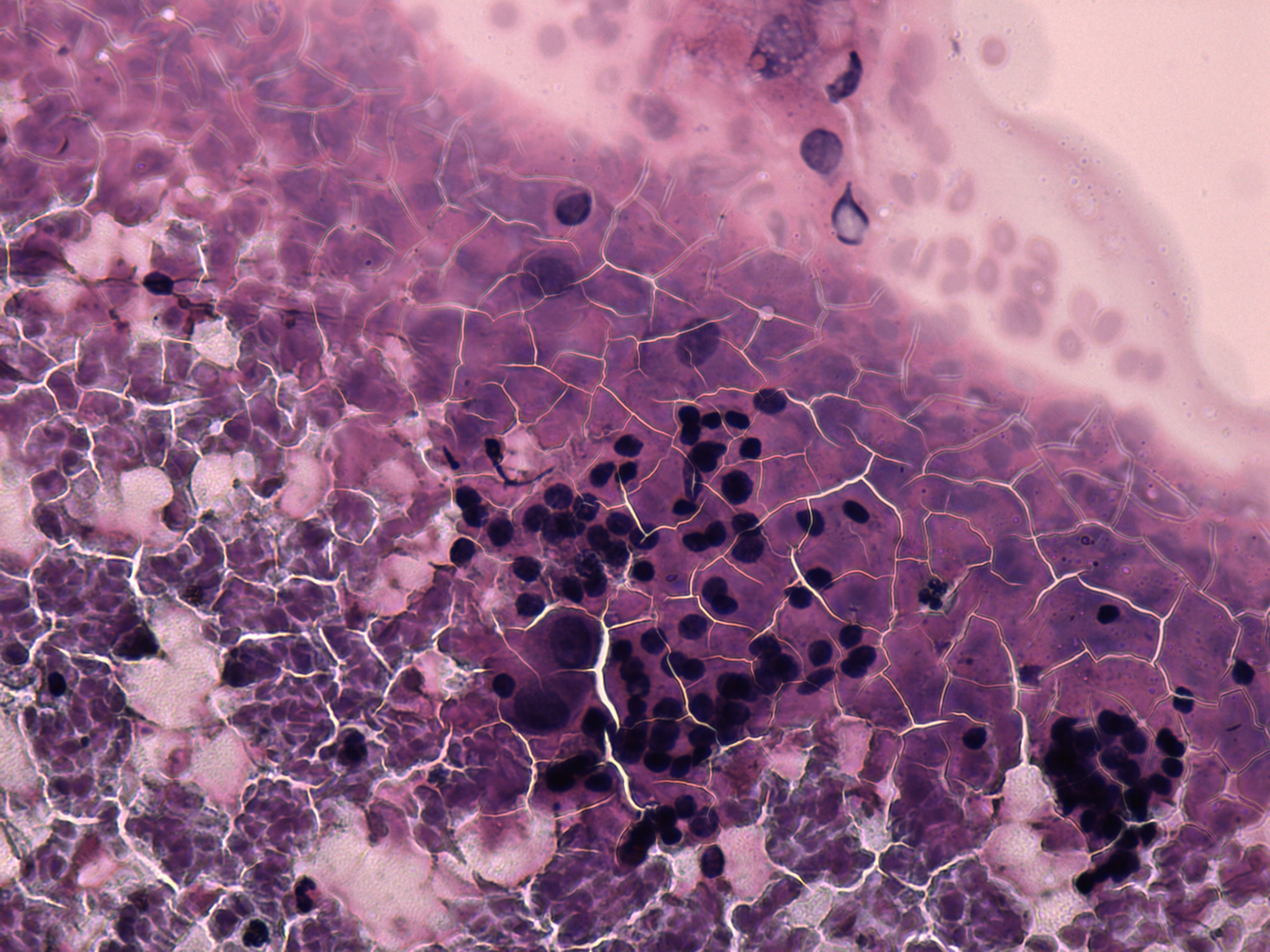
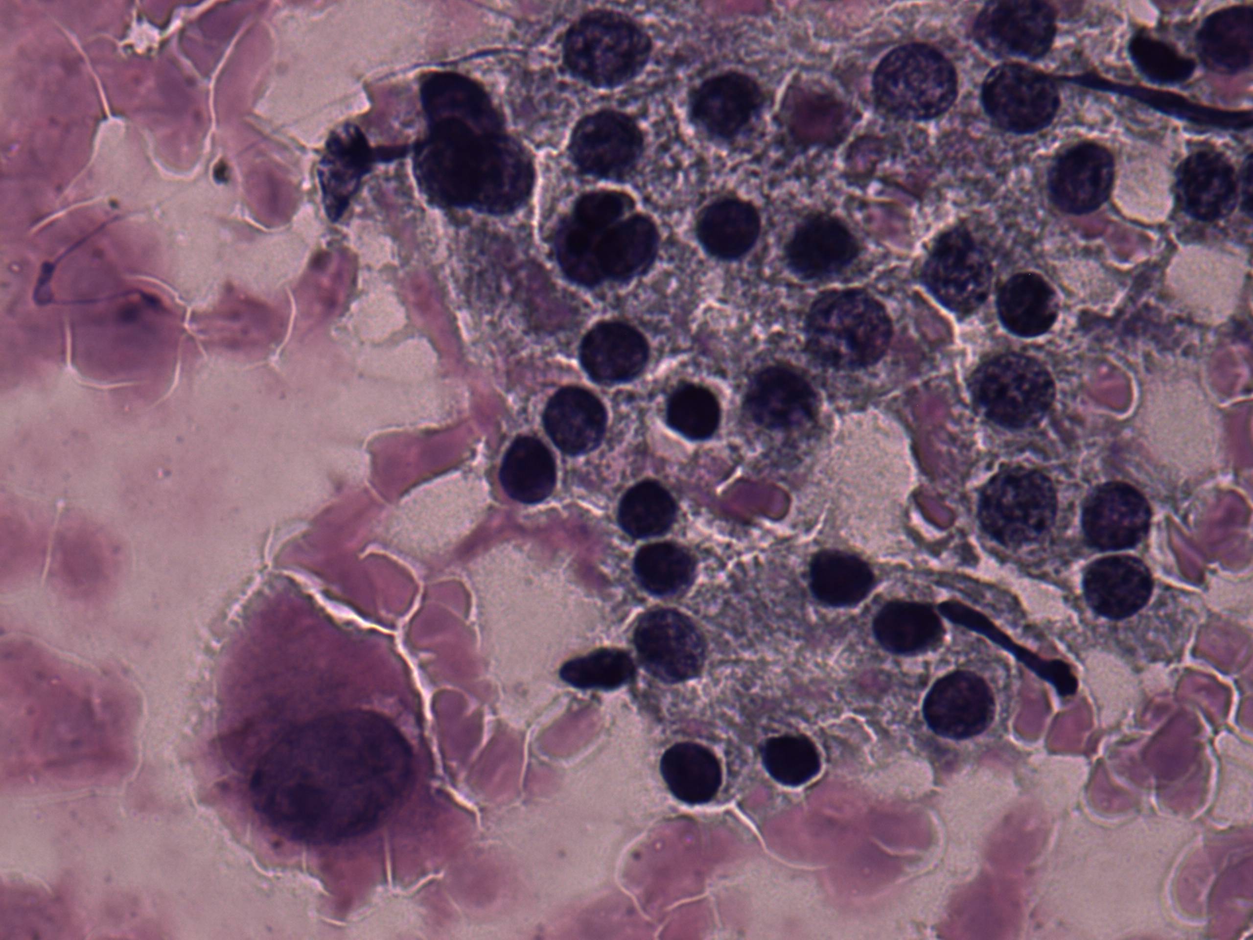
Two cell populations are demonstrated. Firstly, small pycnotic nuclei
which predominate the smear. Secondly, there are a few enlarged
atypical cells with abundant cytoplasm and prominent nucleoli.
|
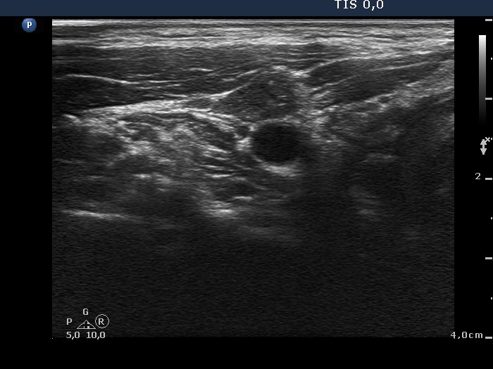
Ultrasonography of the right thyroid
|
 |
 |
| The sonographic pattern is suspicious
because the nodule contains hyperechogenic granules and has blurred
borders. |
Ultrasonography of the right side of the neck
|
 |
 |
| There is enlarged lymph node
containing microcalcifications and lacking regular hilum. |
|
The cytological findings were not enough to raise the possibility of
malignancy, they corresponded to AUS category of Bethesda system.
Taking the sonographic pattern into account, a combined
sonographic-cytological diagnosis could raise the possibility of a
thyroid carcinoma. It is worth mentioning that 9 aspirations occurred
in this patient at two different occasions and only one smear contained
a few suspicious cells. It seems possible that if we followed the
Bethesda protocol, atypical cells would be missed later in the course
and longer delay would occur in the correct therapy.
|
