|
|
 |
 |
|
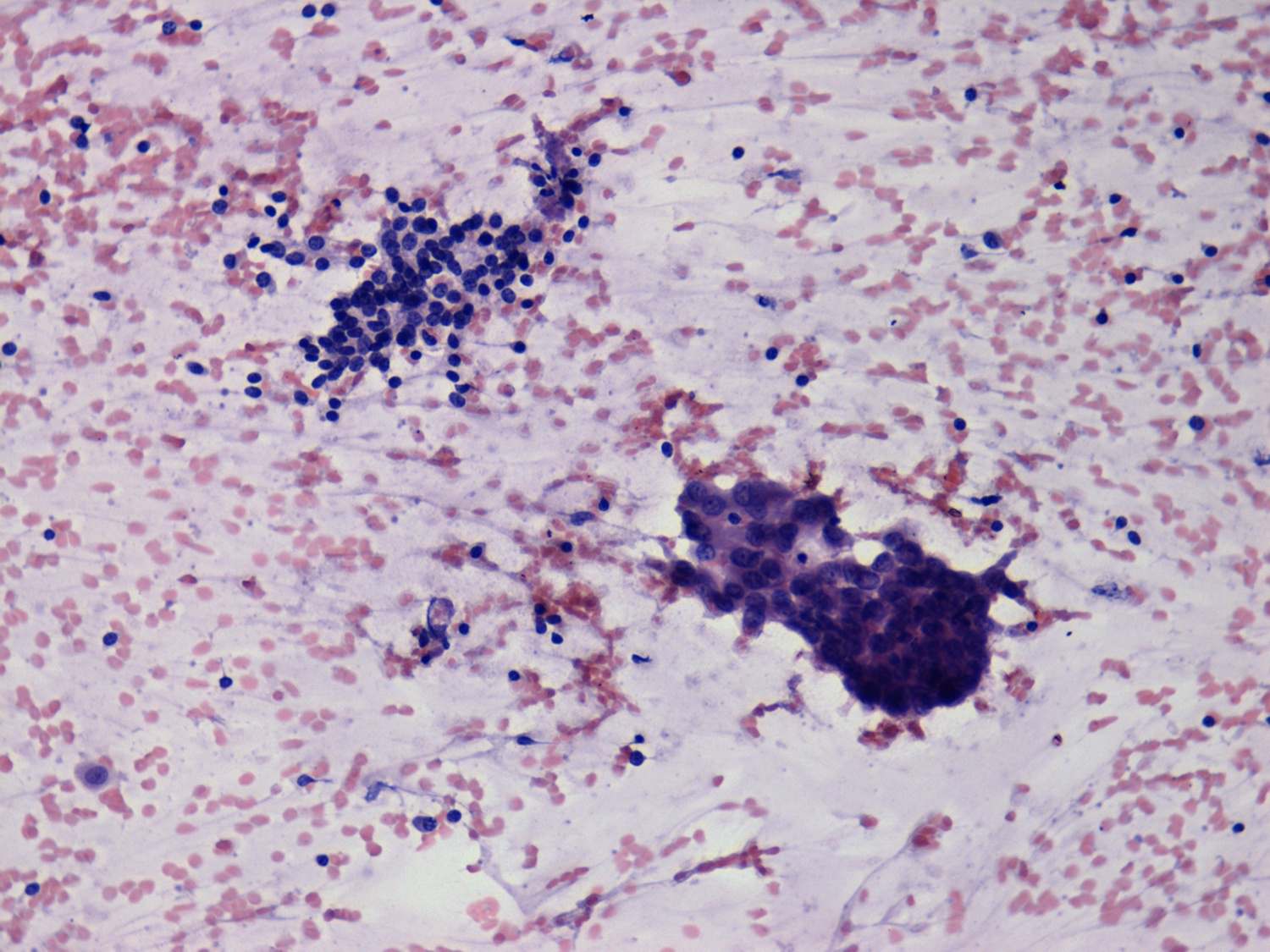
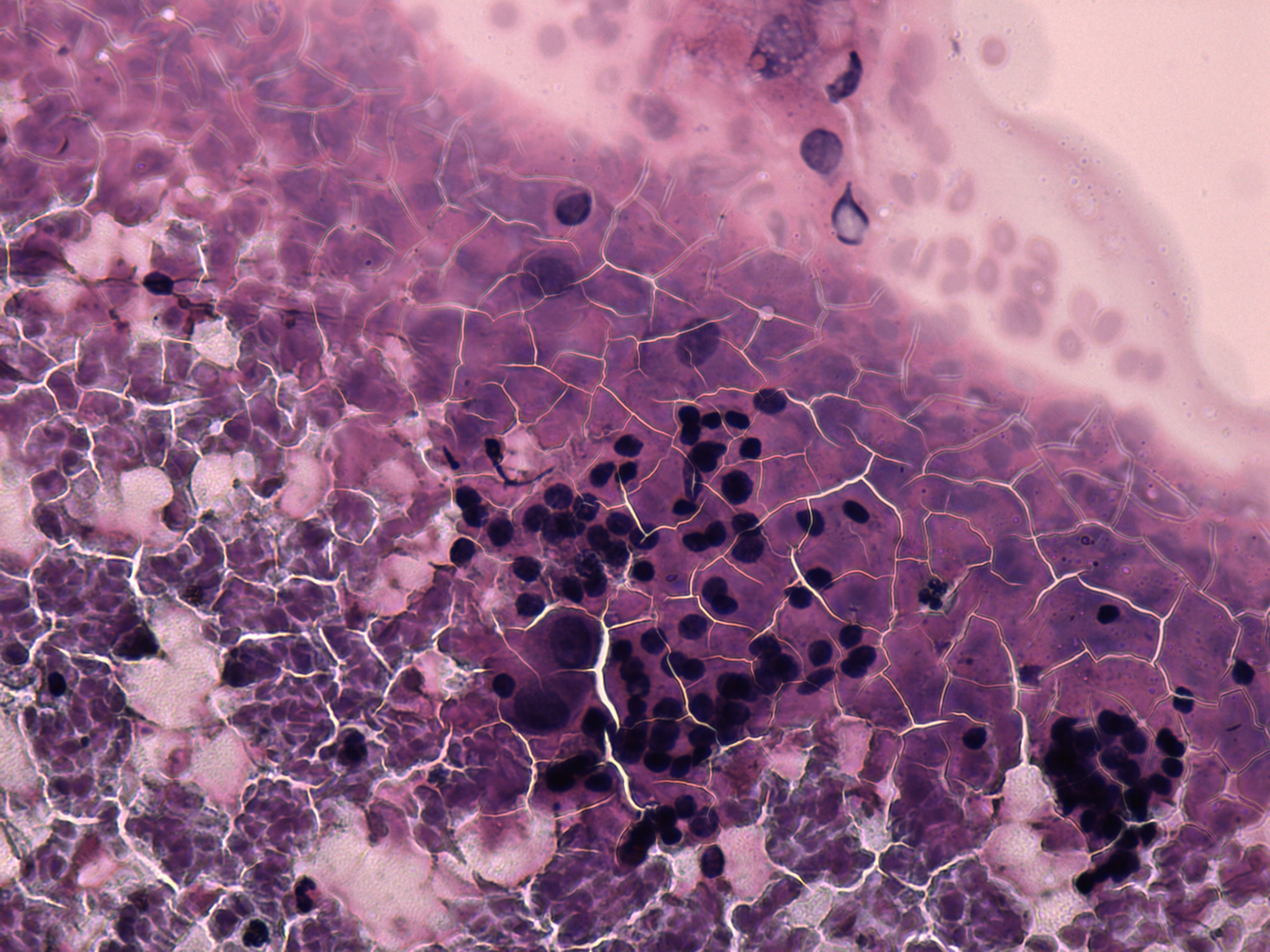
Benign appearing cells in the upper part while a suspicious papillary
group in the lower part of the image.
|
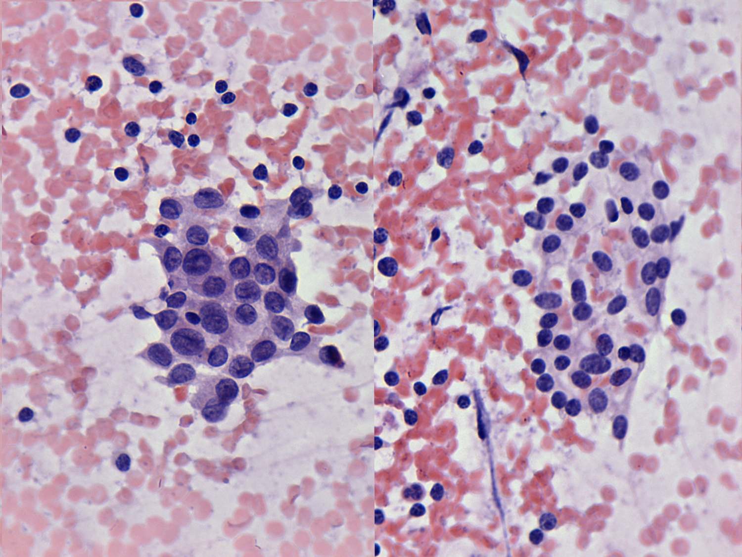
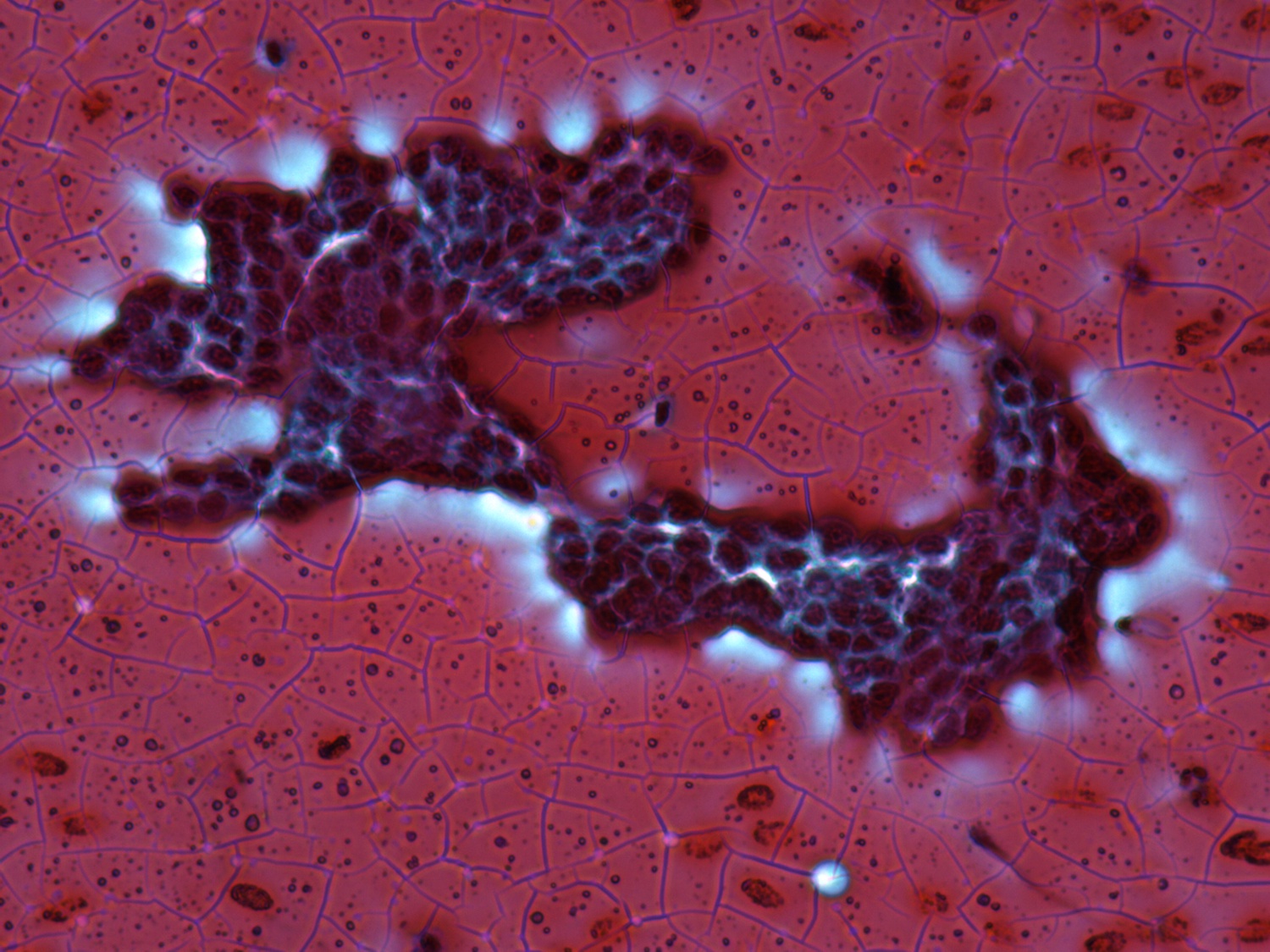
Left suspicious, right benign cell cluster. |
| |
|
|
 |
 |
 |
 |
|
Two cell populations are demonstrated. Firstly, small pycnotic nuclei
which predominate the smear. Secondly, there are a few enlarged
atypical cells with abundant cytoplasm and prominent nucleoli.
|
| |
|
|
 |
 |
|
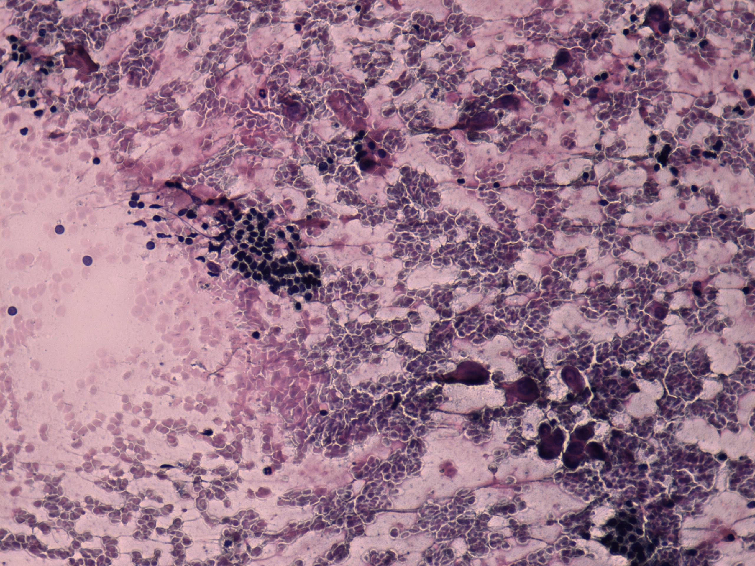
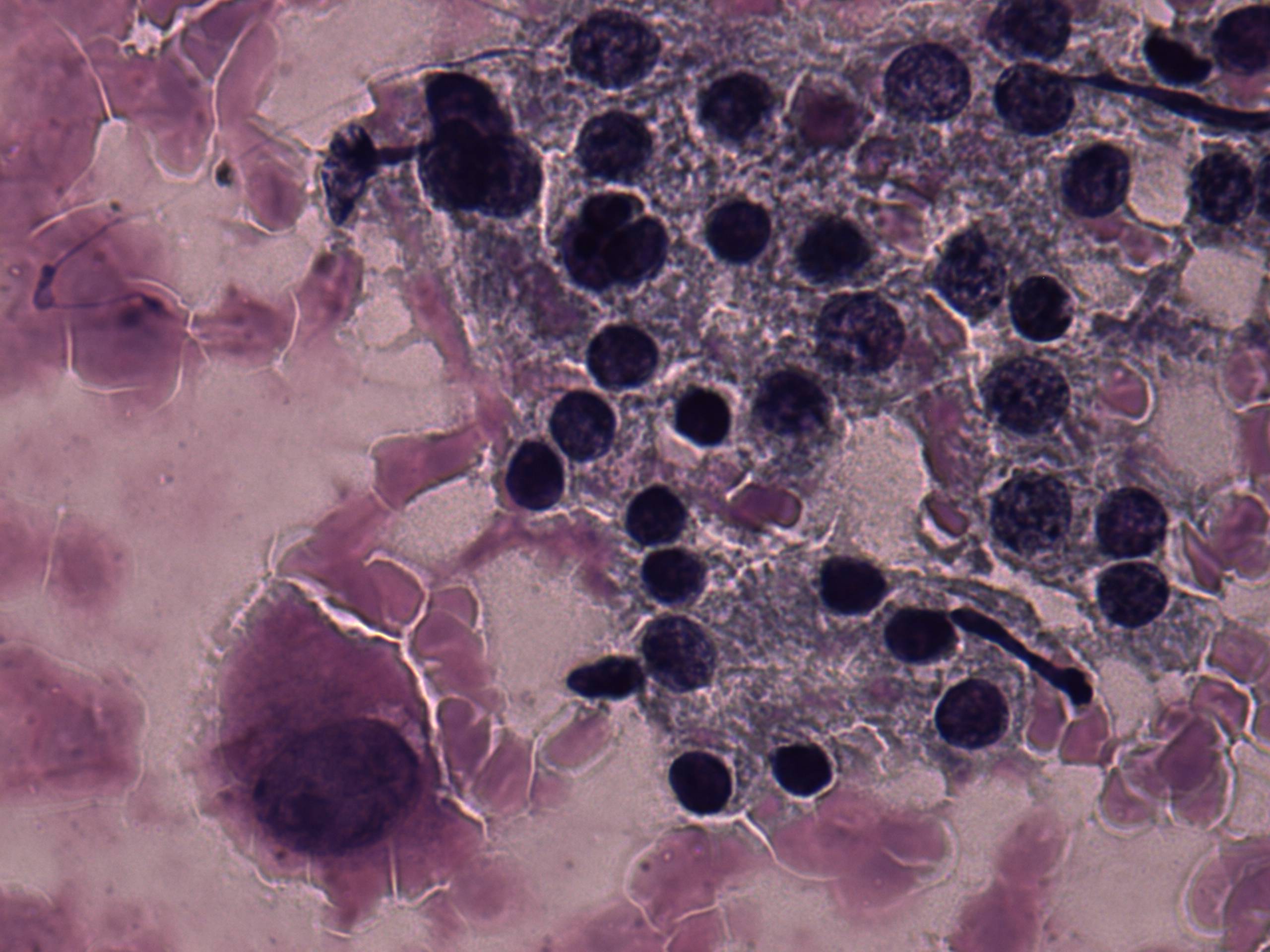
The nuclear details of the papillary cluster is difficult to judge
because the nuclei are pycnotic, but this papillary cluster corresponds
to a hyperplastic one because of relatively sharp edges an peripheral
vacuolization.
|
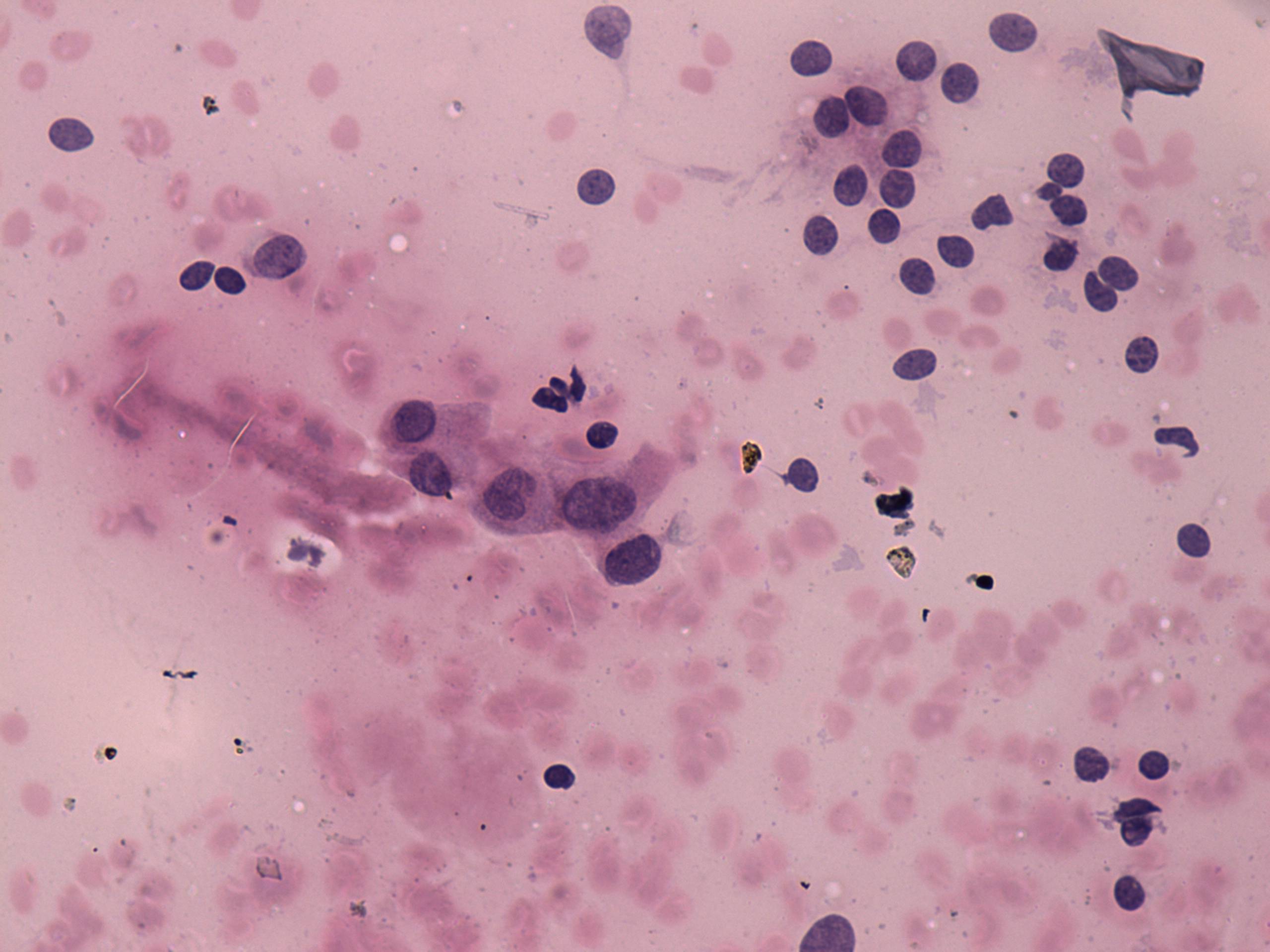
These monolayered sheets are composed of suspicious cells because of
the presence of intranuclear inclusions.
|
