|
|
 |
 |
 |
 |
|
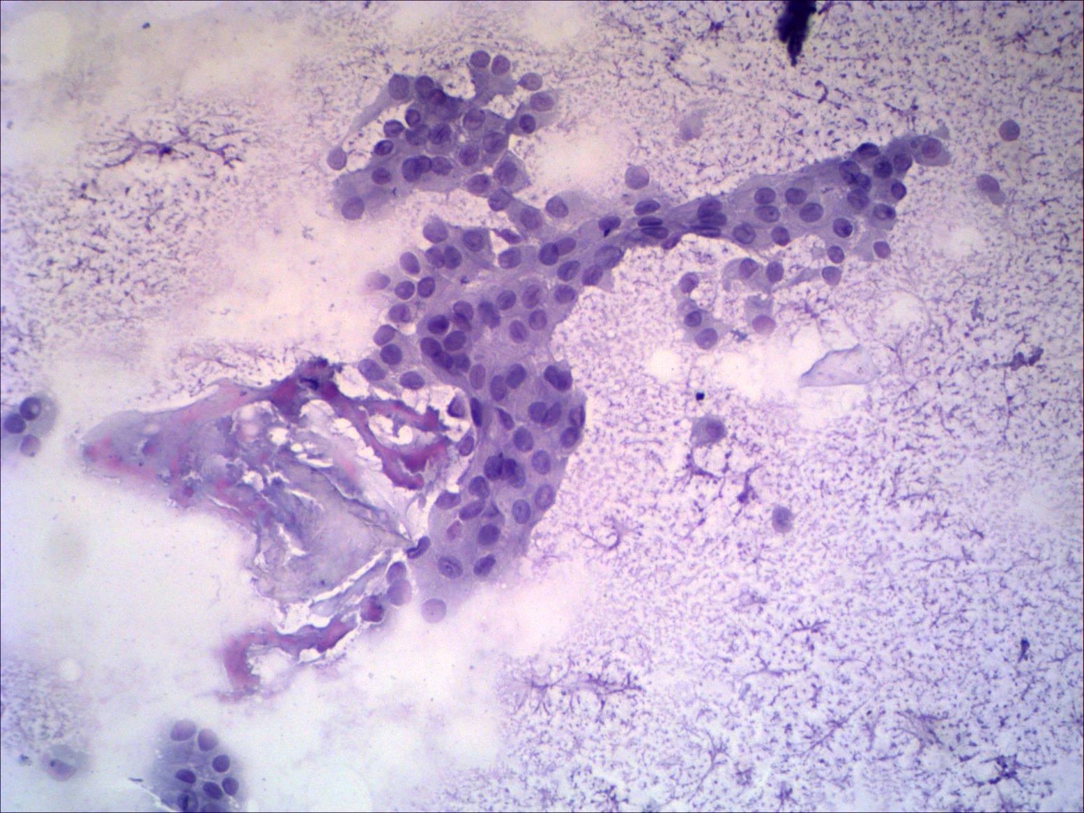
Monolayered sheets of cells in difuuse colloidal background. Note loss
of polarity. The significance of large number of grooves is limted
because follicular cells exhibit signs of oxyphilic metaplasia. The
interpretation of the intranuclear hole in the right lower image is
ambiguous because it is not surrounded with dark sharp ring. Taking all
these into account an oxyphilic tumor has to be considered.
|
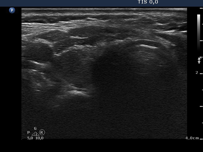
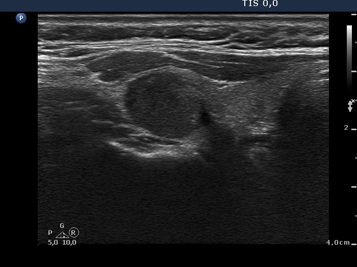
Ultrasonography of the right thyroid
|
 |
 |
|
The sonographic pattern is suspicious because the nodule has irregular
borders. The lack of halo and perinodular blood flow are strong
arguments against a follicular type tumor, i.e. Hürthle-cell
adenoma.
|
|
The cytological findings was not enough to raise the possibility of a
papillary carcinoma but combining the cytological and sonographic
properties we gave a common cytological-sonographic diagnosis of
suspicion of papillary cancer.
|
