|
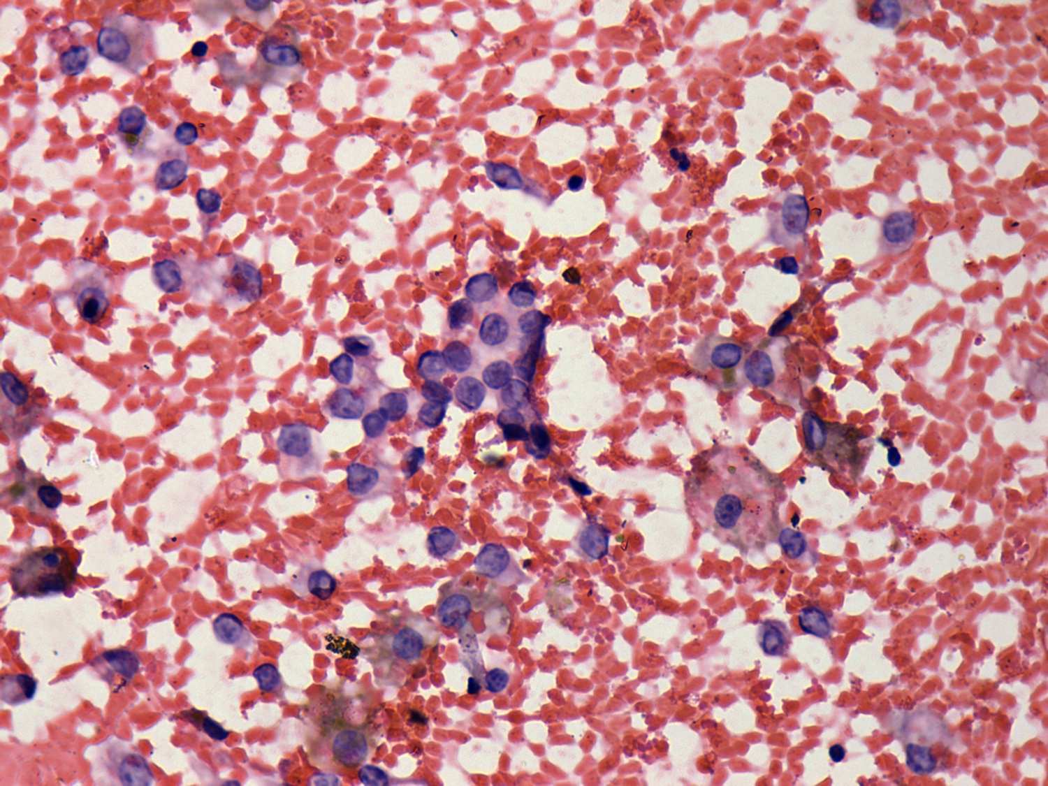
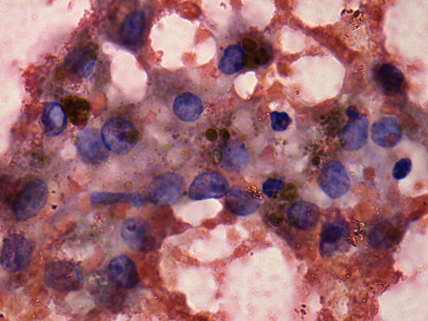
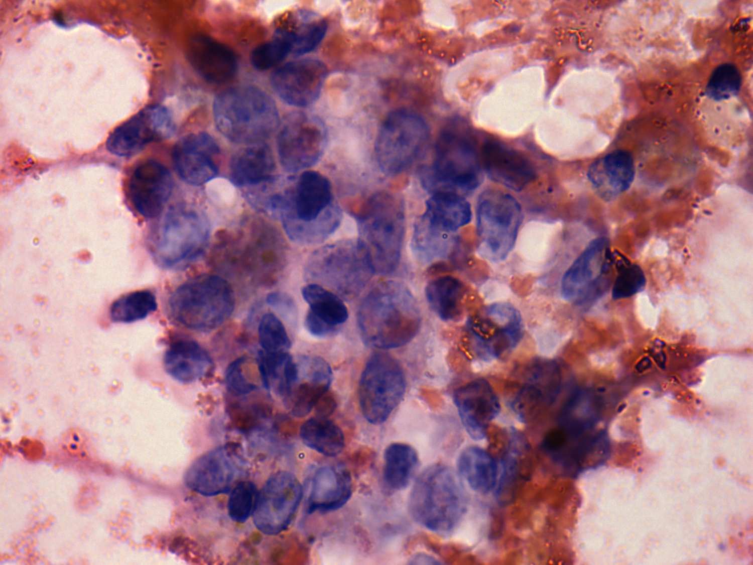
The smear was prepared from cystic fluid. The disarranged structure
presented on the right lower image was not reassuring. The inclusion
demonstrated in left lower image must not to overrate because of its
irregular shape and the presence of degenerative changes. Nevertheless,
in the case of a cystically degenerated papillary carcinoma we can
hardly await a classical cytological presentation.
|