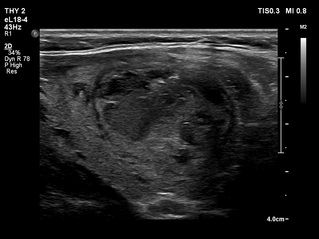
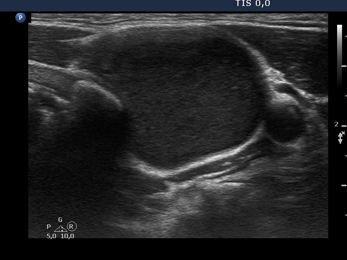
PAPILLON COURSE on THYROID ULTRASOUND

Section 2 The nodular goiter

Part 2 Composition of the nodule
Lectures


Table 4 Technical issues

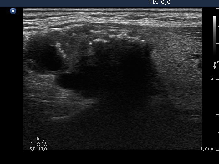
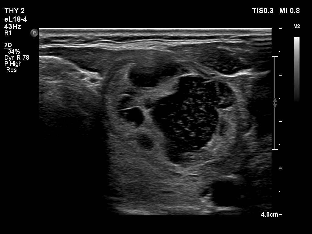
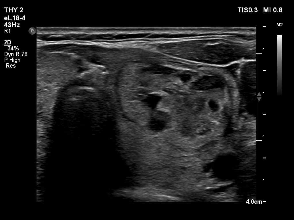
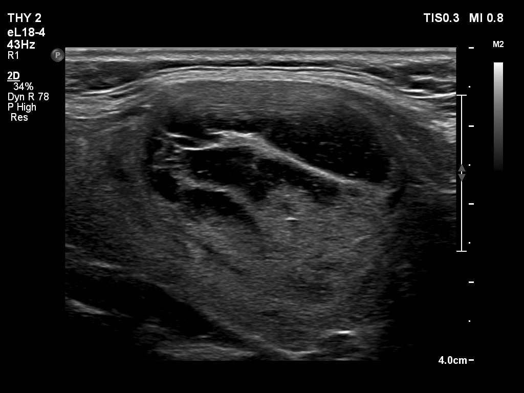
Here we discuss some frequently occurring technical problems. These mainly rely on the influence of the fluid either on the presentation of the nodule as a whole or on the solid part. Another issue is that the cystic fluid occasionally seems to be not anechoic.


Papillary carcinoma (histology) - case conp044

An acoustic shadow hinders the judgement of the dorsal portion of the nodule. This part of the lesion is anechoic similarly to a cystic area. This tumor had cystic parts marked with red arrows, but the portion between yellow arrows is impossible to judge because of the acoustic shadow.

Benign cystic nodule (cytology) - case 1497
Before the aspiration
After the aspiration

The nodule started refilling during the aspiration (see video record). The moderately hypoechoic mass in the right lower image corresponds to the newly appeared blood. Note that the fresh blood is not echo free.

Benign cystic nodule (cytology) - case 126

The echogenicity of the large hypoechoic mass in the upper 4/5 of the nodule was deceptive. It is not a solid portion but a cystic area. By compressing the nodule with the ultrasound probe, the flow of the mass is clearly visible (see video). Such deceptive presentation of fluid is a rare phenomenon, we meet this e.g. in inflammatory cyst content.

Benign cystic nodule (cytology) - case 2071
First examination
30 months after the first examination

In contrast with the first examination, the echogenicity is not echo free but deeply hypoechoic on follow-up examination. Note the presence of septa.

Benign cystic nodule - case 1275

The aspiration of the cystic content has led to two important changes: the solid part became more hypoechogenic and the borders of the nodule became irregular, spiculated and lobulated. The explanation of the first phenomenon is that the echogenicity increases dorsal to cystic areas.

mask