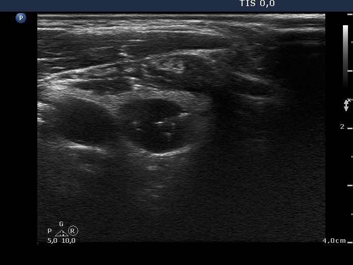
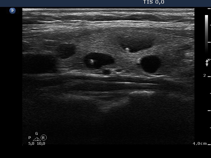
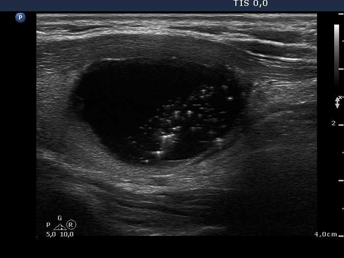
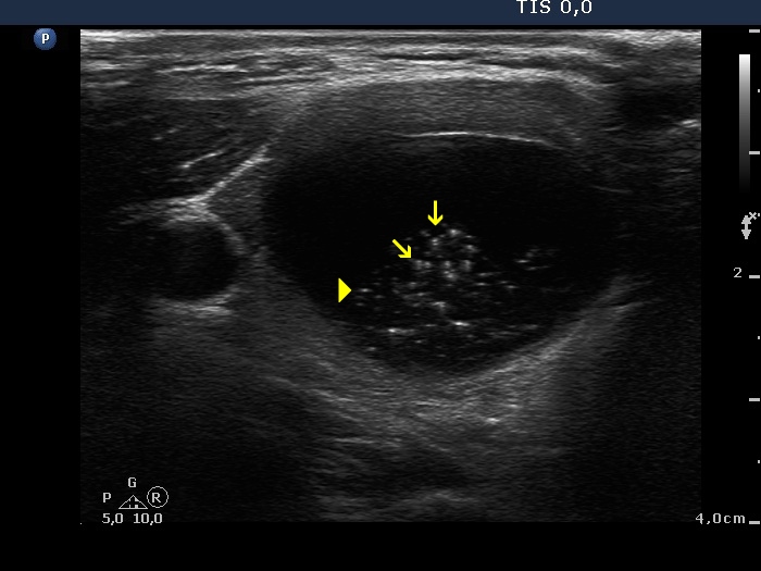
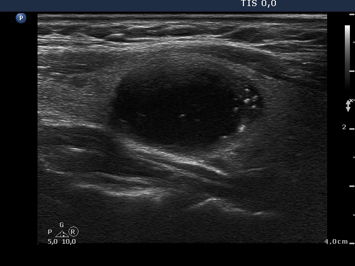
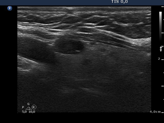
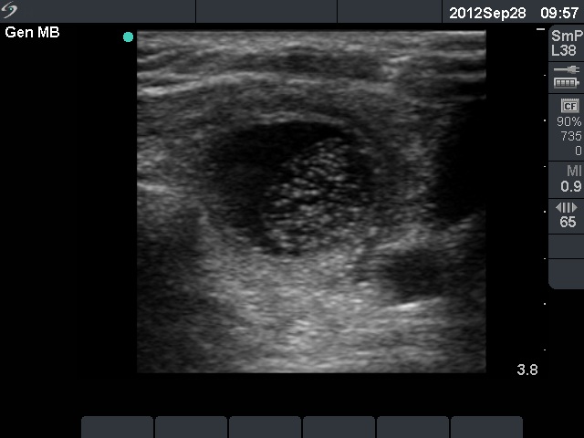
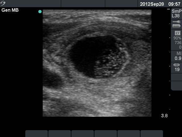
Benign cystic-colloid goiter (cytological diagnosis) - case 284 |
 |
 |
|
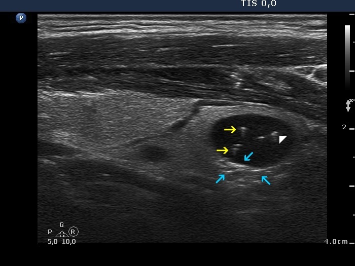 |
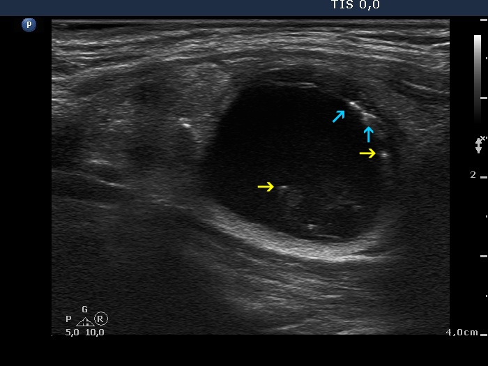
The figure marked with white double arrow is ambiguous because of the lack of dorsal tail. This figure is composed of two granules which might be two punctate echogenic foci. However, there are typical forms of comet-tail artifacts, the presence of which makes it very likely that the equivocal figures are also comet-tail artifacts. Moreover, these are within cystic fluid which theoretically excludes that these might be pathognomic microcalcifications. In addition to it is worth analyzing the dorsal wall of the cyst and the thyroid parenchyma dorsal to the nodule, both have hyperechogenic figures corresponding to a posterior back wall enhancement.
|
| |
|
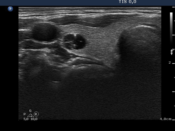
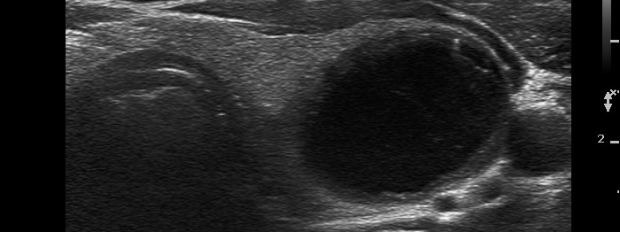
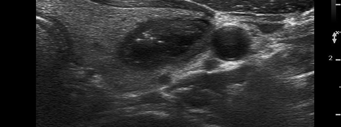
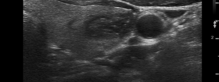
Benign follicular proliferation (cytological diagnosis) - case 270
|
 |
 |
A typical comet-tail artifact is demonstrated. |
| |
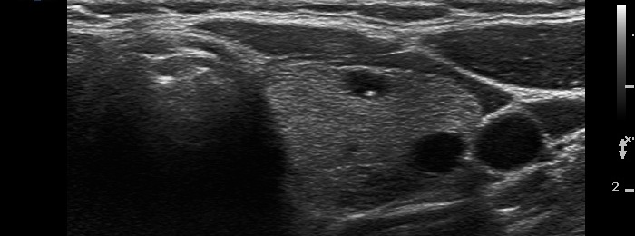
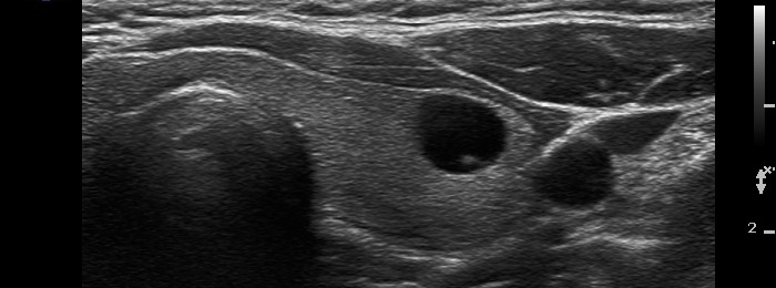
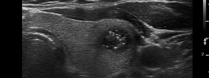
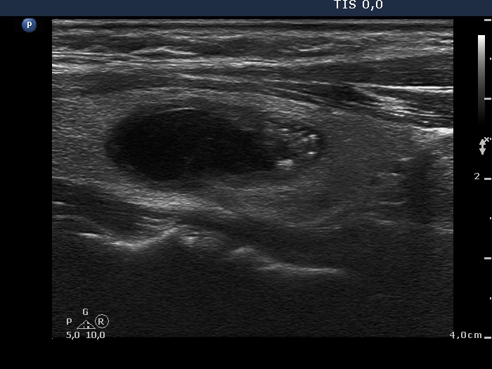
Intact thyroid with cystically dilated macrofolliculi (histological diagnosis) - case 1539
|
 |
 |
Right lobe: there are typical comet-tail artifacts, two in the left horizontal scan and one in the right longitudinal scan. The latter presents two more cystic lesions (the lower two) in which the hyperechogenic figures have only a vestigial dorsal tail. |
  |
 |
Left lobe: there are typical comet-tail artifacts in this lobe, too. |
| |
Benign colloid cyst (cytological diagnosis) - case 402 |
 |
 |
 |
 |
There are several typical comet-tail artifacts in both lobes.
|
| |
Benign cystic degeneration (cytological diagnosis) - case 624 |
 |
 |
 |
 |
|
 |
There are several unambiguous figures with typical presentation (yellow arrows), while several hyperechogenic granules lack dorsal acoustic shadow (white double arrows) which theoretically might be punctate echogenic foci. On the other hand, in the presence of typical comet-tail artifacts, similarly bright non-typical forms more likely belong to the same category. Moreover, punctate echogenic foci (microcalcifications) cannot be found in cystic fluid.
|
| |
Follicular adenoma (histological diagnosis)
|
Before aspiration of 2 mL cystic fluid |
  |
 |
There are numerous hyperechogenic granules in the lower pole of the nodule. These figures might be confused with punctate echogenic foci (microcalcifications), as happened in this case. However, several have dorsal tail and therefore the remaining without a tail likely belong also to the same subgroup.
|
After aspiration of 2 mL cystic fluid |
  |
 |
It became more evident after removal of the cystic fluid that the figures are comet-tail artifacts.
|
| |
|
Hashimoto's thyroiditis with several cystic areas but without any nodules (histological diagnosis) - case 1365
|
 |
 |
In this case we found only hyperechogenic lines. This is an unusual presentation of a comet-tail artifact. The video is clearly superior to a saved image, in the former the hyperechogenic figure appeared in a more typical form.
|
| |
|
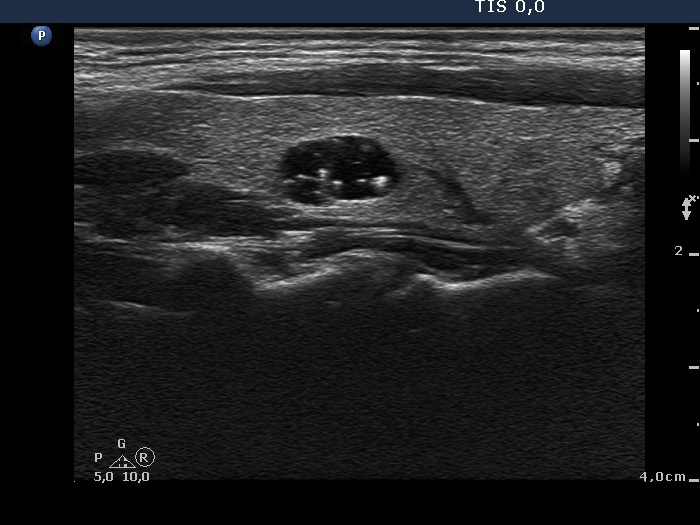
Benign colloid goiter (cytological diagnosis) - case 386 |
 |
 |
At first sight, this pattern mimics the starry sky phenomenon which would be caused by numerous microcalcifications. However, these granules are found in a cystic fluid therefore they cannot be punctate echogenic foci (microcalcifications).
|
| |
|
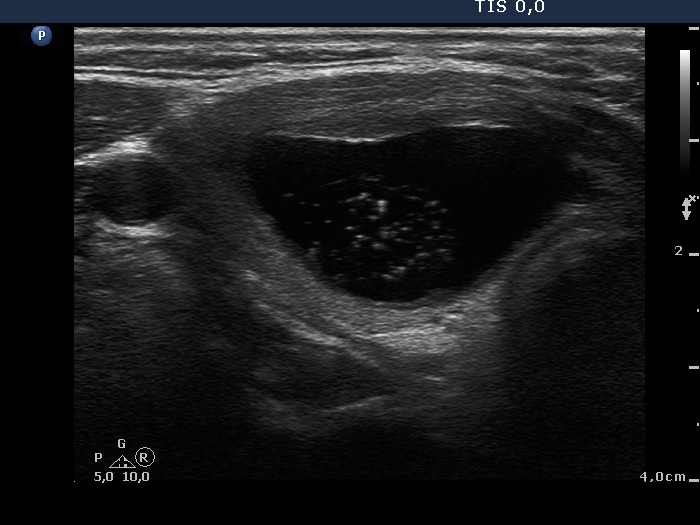
Benign cystic degeneration (cytological diagnosis) - case 662
|
 |
 |
It is worth analyzing the hyperechogenic figures in the central part of the cystic area. One in the horizontal view and another one in the longitudinal scan have a broader than usually fading tail. The figures pointed with arrows at the border of the cystic and solid part (right side of the nodule in the right image) are posterior back wall enhancement caused by the microcystic area ventral to them.
|
| |
|
| |
|
| |
|
| |
|
| |
|
