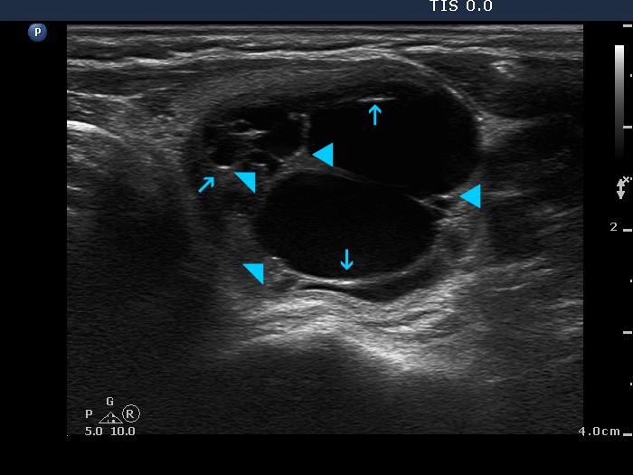
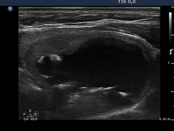
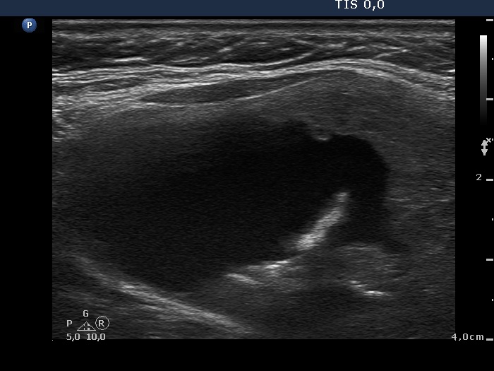
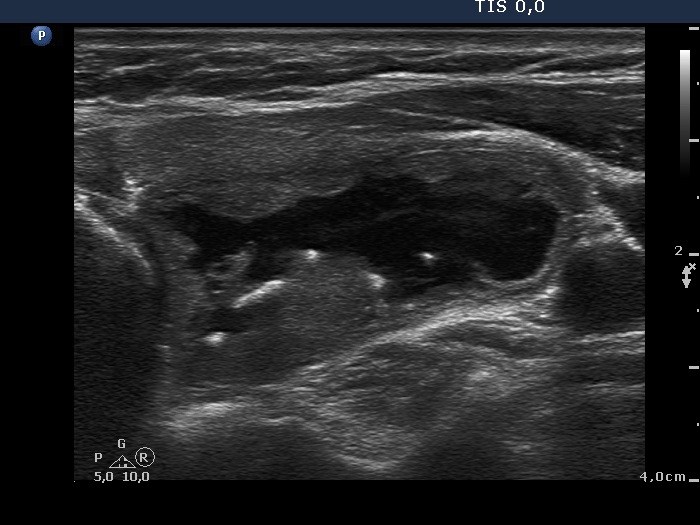
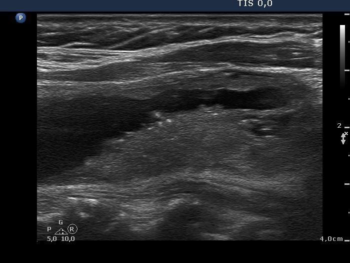
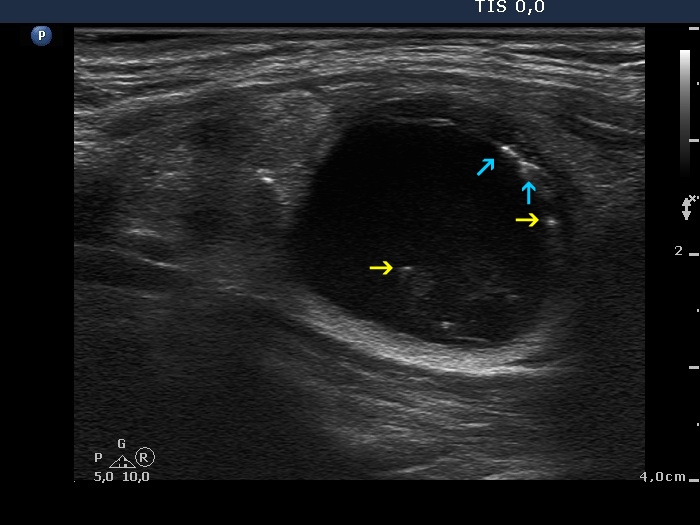
Selected topics - intranodular hyperechogenic figures - Table 3. Cystic back wall figures

doi: 10.24390/thyrosite.2018.ihf.03.1

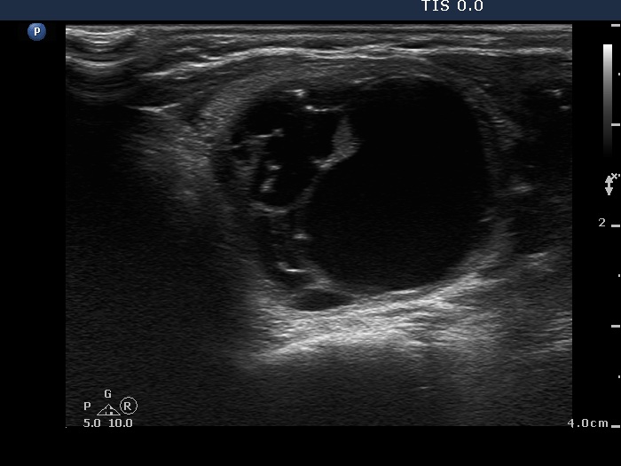
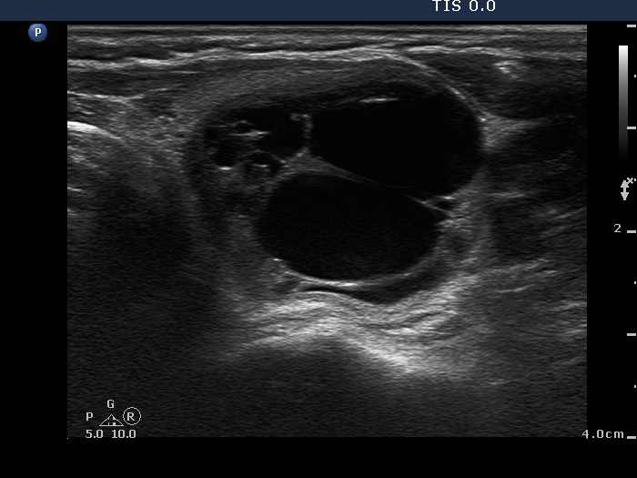
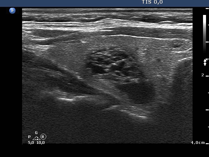
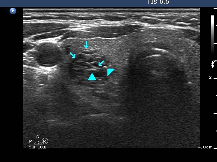
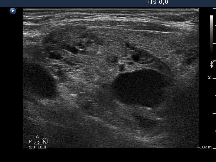
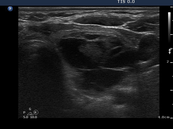
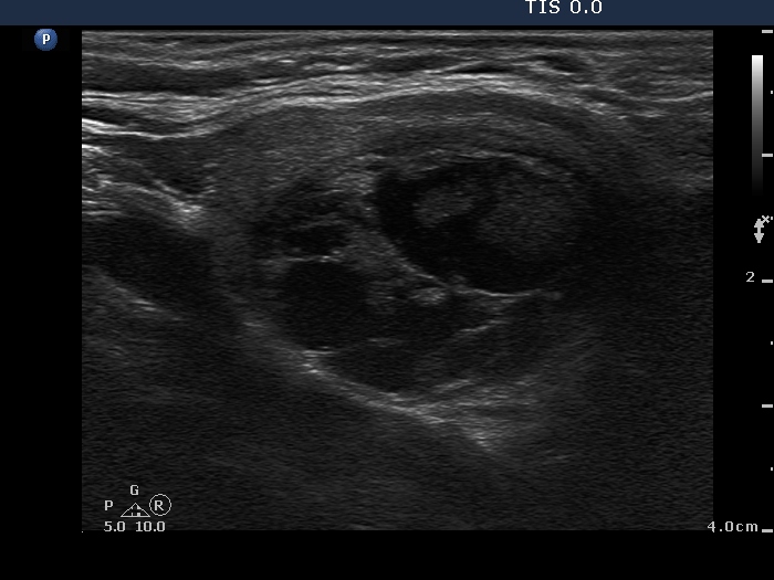
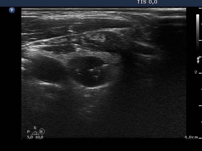
A punctate echogenic focus is a round bright granule. The size of this figure is less than 1 mm, usually is around 0.5 mm. A lesion which has typical punctate echogenic foci (microcalcifications) almost always presents less bright granules, too. The latter might be non-typical forms of microcalcifications or non-specific granulations. In fact the identification of punctate echogenic foci (including microcalcifications) is a matter of exclusion other forms of hyperechogenic figures, i..e comet-tail artifacts, connective tissue and posterior back wall enhancement. All of the latter have additional features lacking in the event of punctate echogenic foci. Nevertheless, it means that atypical presentations of these figures might mimic punctate echogenic foci including microcalcifications.
The presentation of punctate echogenic foci depends highly on the ultrasound device. If the resolution is worse than the granule is larger (see the lowest image). On the other hand there is a very harmful trend in newer equipments which might cause difficulty in recognition of punctate echogenic foci (microcalcifications). This trend is the "overharmonization" which means that the normal grainy structure of the thyroid is masked in order to gain a more harmonic, beautiful image. It has practically no sense, moreover the overharmonization masks the pathologic hyperechogenic granules, the microcalcifications, too, which is a very serious hazard.
We prefer the term punctate echogenic foci instead of microcalcification in order to avoid misnomer for granules caused by other pathological structures. Nevertheless, in many publications and even in other parts of this website we use the term "microcalcification" for every punctate echogenic foci.

Benign cystic lesion (cytological diagnosis) - case 1139

The posterior acoustic enhancement in the dorsal wall of the small cystic areas causes no concern if this optical artifact is linear (arrows), however granular figures might be misinterpreted as punctate echogenic foci (microcalcifications) (arrowheads).


Benign cystic lesion (cytological diagnosis) - case 28

The hyperechogenic lines in the dorsal wall of the cystic area do not cause concern (arrows), while the granules (arrowheads) might do.


Benign hyperplastic nodule (histological diagnosis) - case 653

Note the linear figures in the back wall of the cystic areas. The ventral solid area of the nodules contains both hyperechogenic lines and granules; therefore these figures correspond to connective tissue. It is ambiguous whether the echogenic figures in the dorsal solid area (right image) are presentations of connective tissue or posterior acoustic enhancements but the former seems to be more likely.


Follicular adenoma (histological diagnosis) - case 486

The dorsal wall of the small cystic areas presents posterior back wall enhancement. The origin of the large granules within the solid part remains obscure; nevertheless these are probably colloid crystals.


Follicular adenoma (histological diagnosis) - case 108

Almost every cystic chamber presents the back wall posterior enhancement.
   

Benign cystic lesion (cytological diagnosis) - case 284

The cystic part of the lesion contains typical comet-tail artifacts and back wall posterior enhancement. Regarding the latter, note that the hyperechogenic figures appear not only in the dorsal wall of the cyst but even by the dorsal to the back wall in the solid parenchyma.


Benign cystic lesion (cytological diagnosis) - case 420

The lesion presents various forms of hyperechogenic figures including a typical comet-tail artifact (lower left image), a large cluster in the ventral part of the cystic lesion (upper left image) which is probably a large colloid crystal, too. The figures in the dorsal wall are very likely caused by a posterior enhancement.


Benign cystic degeneration (cytological diagnosis) - case 662

The bright figures marked with arrows at the border of the cystic and solid part (right side of the nodule in the right image) can be easily misinterpreted as punctate echogenic foci (microcalcifications); these are posterior back wall enhancements caused by the microcystic area ventral to them.
There are comet-tail artifacts within the cystic fluid with a broader than usual tail.

   
mask