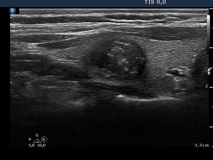
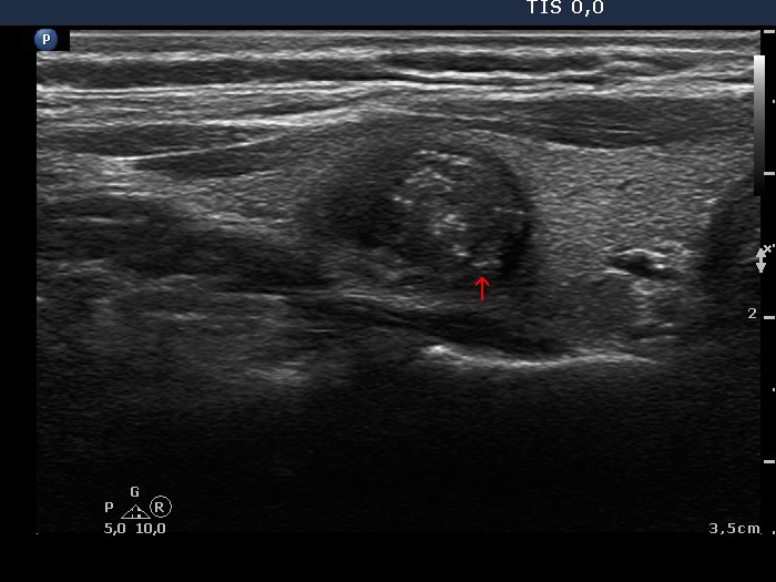
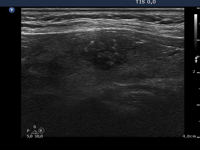
Papillary carcinoma (histological diagnosis) - case 763
|
 |
 |
There are at least 6 bright granules in the left and in the right image. The lesion as most nodules contains non-specific granules and lines which are less bright than the former. The granules of intermediate brightness might be punctate echogenic foci, as well. |
| |
|
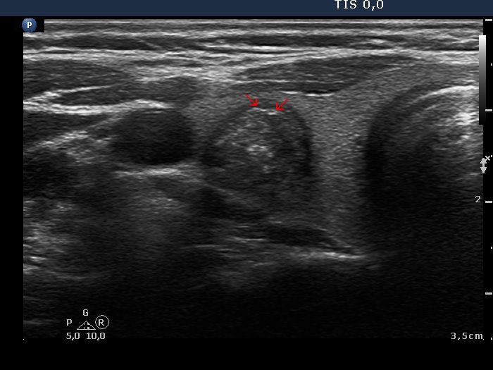
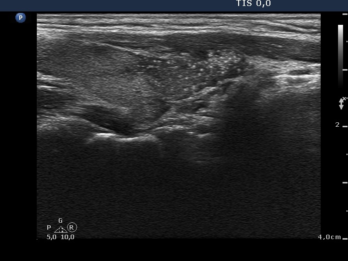
Papillary carcinoma (histological diagnosis) |
 |
|
 |
 |
This case is less edifying or may be more edifying. Compared with the previous case, the granules here are less bright. Nevertheless, great proportion of them belongs to punctate echogenic foci (arrows). It is worth comparing these with non-specific granules (arrowheads).
|
| |
|
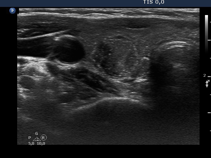
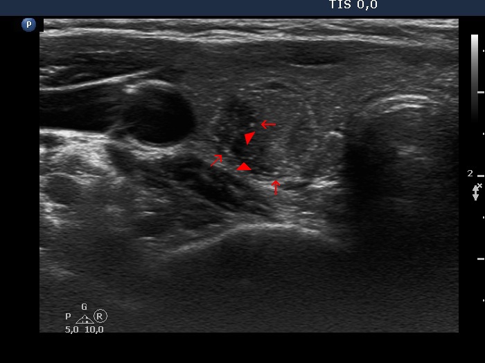
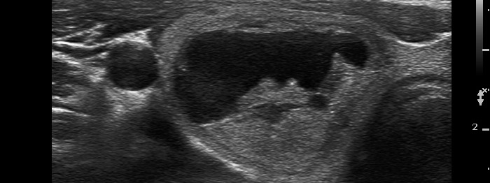
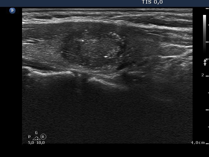
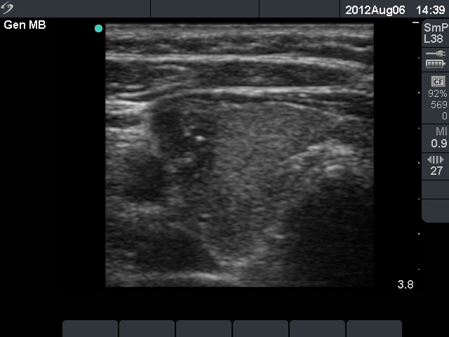
Papillary carcinoma (histological diagnosis) - case 607 |
 |
 |
 |
 |
Arrowheads point to several non-specific granules while arrows do to punctate echogenic foci.
|
| |
|
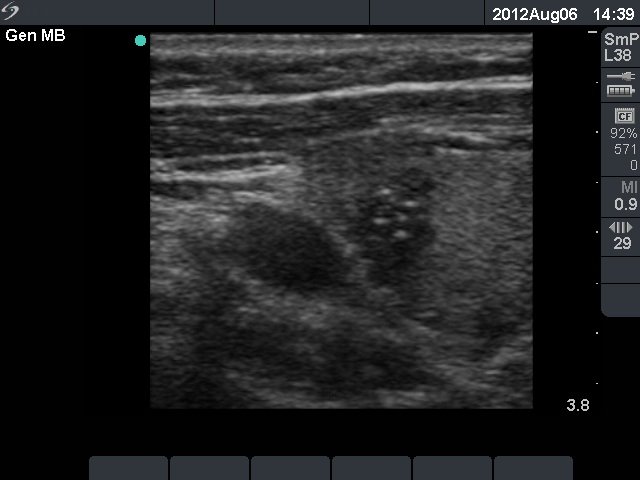
Benign hyperplastic nodule (histological diagnosis) |
Upper and lower horizontal views |
Longitudinal view |
  |
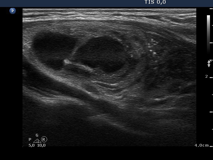 |
The bright granules cannot be categorized other than punctate echogenic foci (microcalcifications). The presentation in the right image is very close to the starry sky phenomenon. Fortunately, such pattern is very rarely seen in benign lesions.
|
| |
|
Benign cystic-colloid goiter (cytological diagnosis) |
 |
 |
 |
 |
This is an ambiguous pattern: although hyperechogenic bright granules dominate the lesion, a few similarly bright figures seem to be linear (arrows). However, two of them are in fact figures composed of two granules (in the left image).
|
| |
|
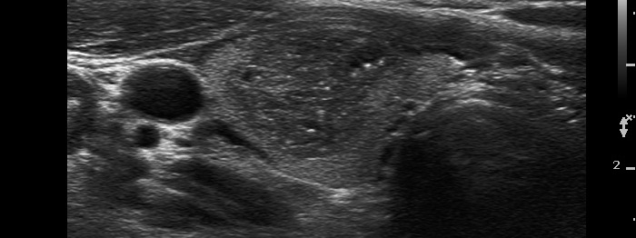
Papillary carcinoma (histological diagnosis) - case p061 |
 |
 |
There is only one granule suspicious being a punctate echogenic focus (microcalcification) in the left, horizontal view while the right, longitudinal view has more typical forms.
|
| |
|
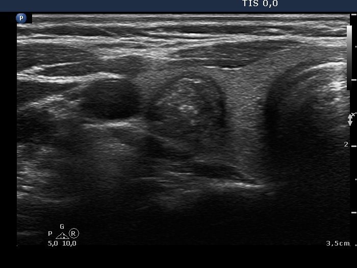
Papillary carcinoma (histological diagnosis) - case 469 |
 |
 |
This tumor contained numerous punctate echogenic foci (microcalcifications). |
| |
|
Hashimoto's thyroiditis (cytological diagnosis) - case 1137 |
 |
 |
A solitary punctate echogenic focus is presented in the right image. |
| |
|
Papillary carcinoma (histological diagnosis) - case p009 |
 |
 |
On histopathological analysis numerous psammoma bodies were found in this tumor. However, the ultrasound presentation is not very convincing. This case illustrates that such pale granules might also be microcalcifications.
|
| |
|
Papillary carcinoma (histological diagnosis) - case 882 |
 |
 |
Smaller and larger bright granules correspond to punctate echogenic foci, in this case to microcalcifications.
|
| |
|
Papillary carcinoma (histological diagnosis) - case 684 |
 |
 |
This examination was performed by older equipment with worse resolution; therefore the granules are not only larger but a bit blurred compared to punctate echogenic foci presented in the former cases.
|
