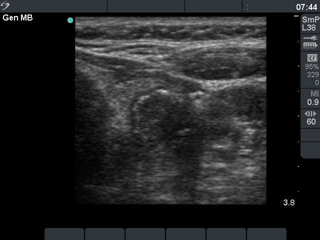
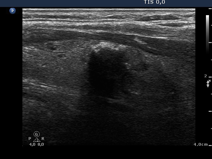
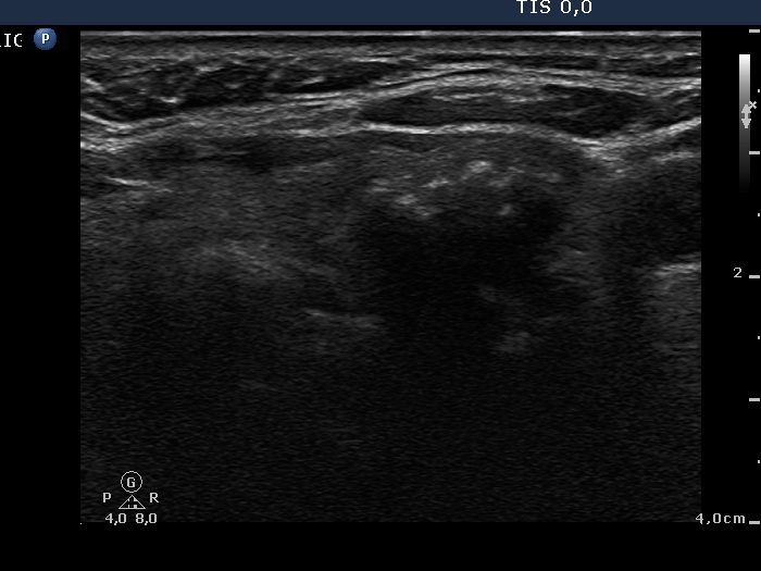
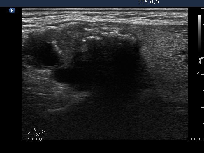
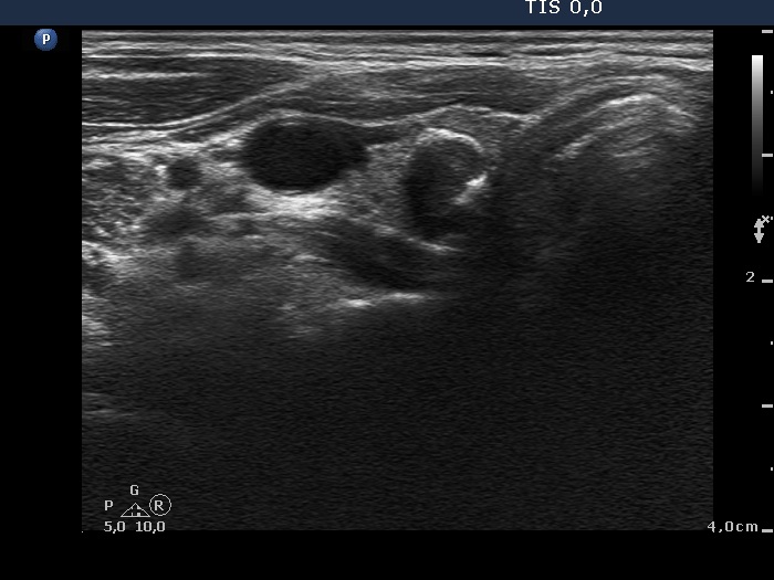
Benign hyperplastic nodule (histological diagnosis) - case 80
|
 |
 |
There were multiple foci of coarse calcification in this case. Note that acoustic shadowing is complete only at the edges of the lesions.
|
| |
|
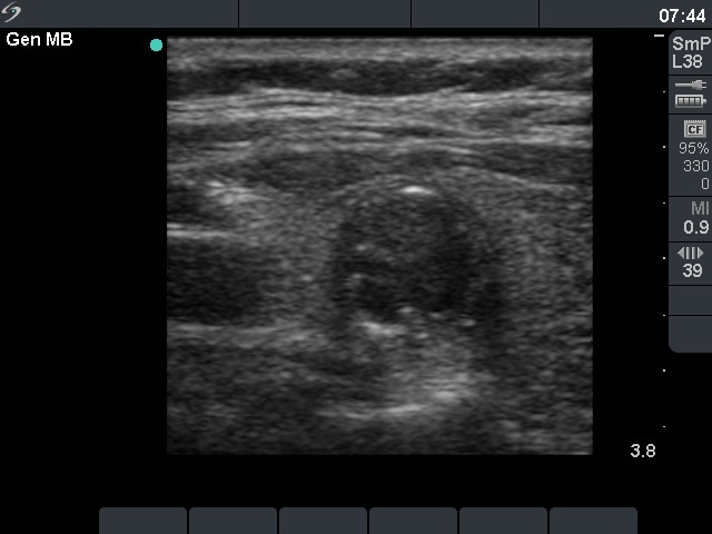
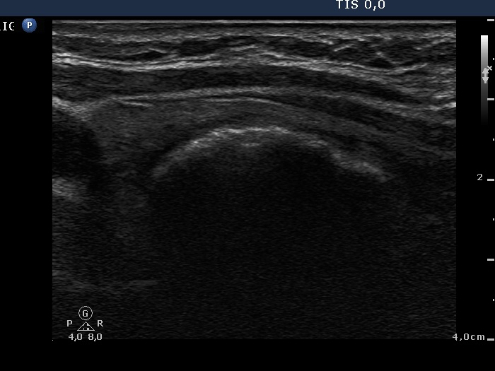
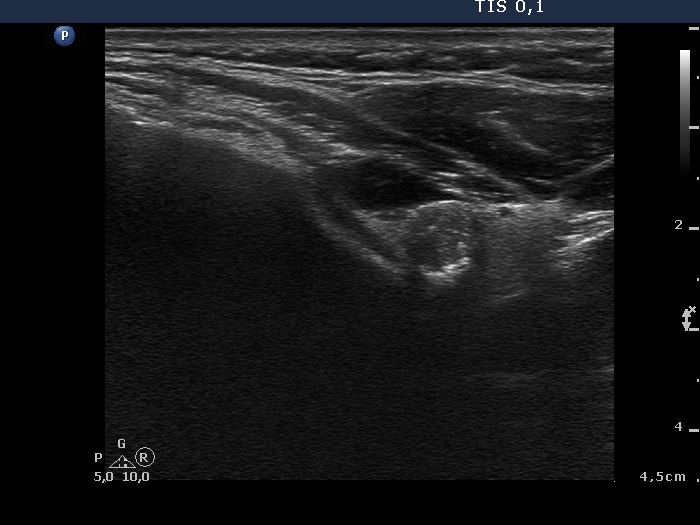
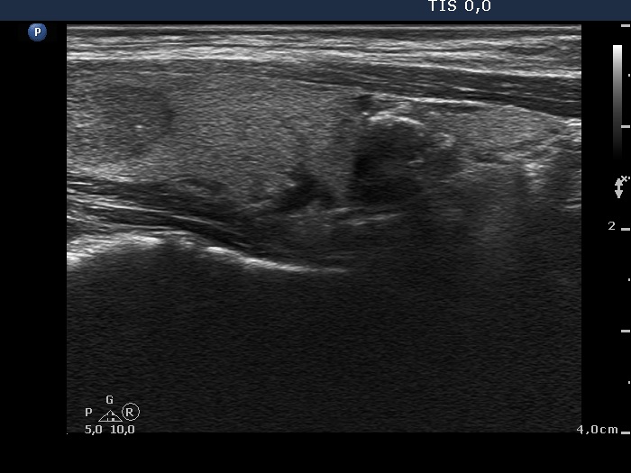
Benign hyperplastic nodule (histological diagnosis) - case 489 |
 |
 |
The nodule had an eggshell calcification.
|
| |
|
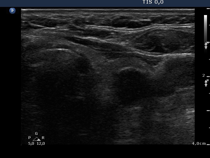
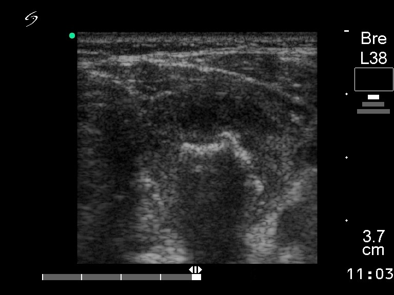
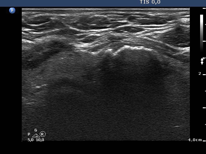
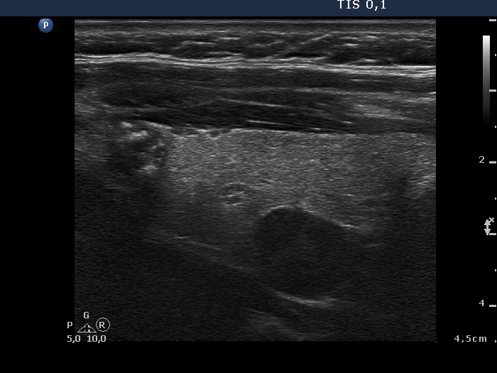
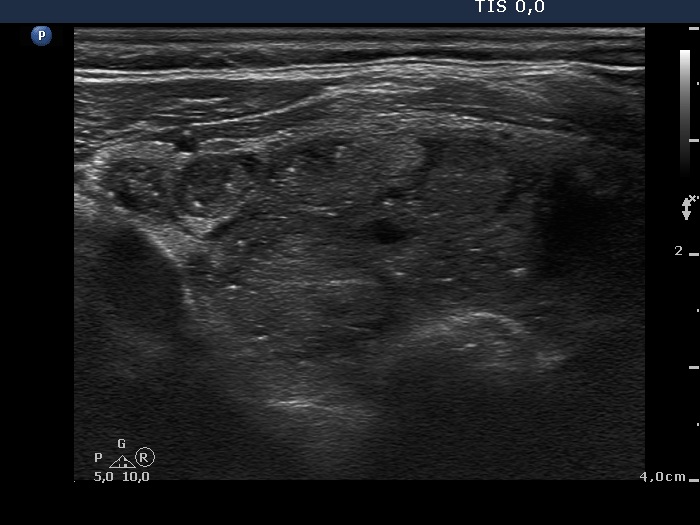
Benign colloid goiter (cytological diagnosis)
|
 |
 |
This nodule presents different types of hyperechogenic granules, the ventral figures are either comet-tail artifacts or punctate echogenic foci, while although the primary focus cannot be seen, the acoustic shadows prove that there are coarse calcifications, as well.
|
| |
|
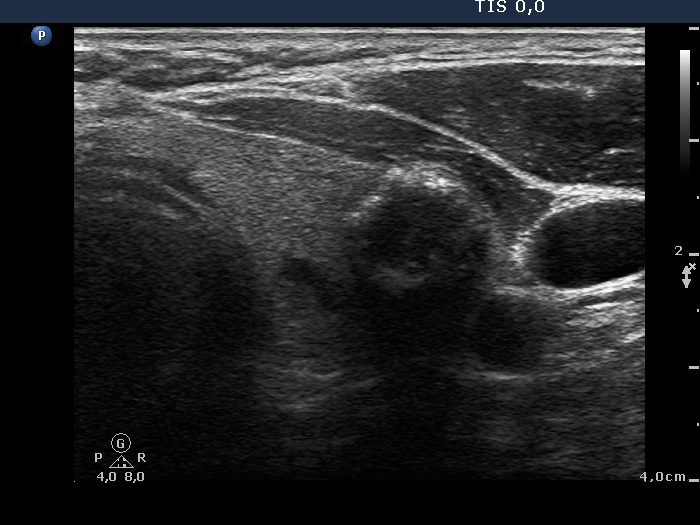
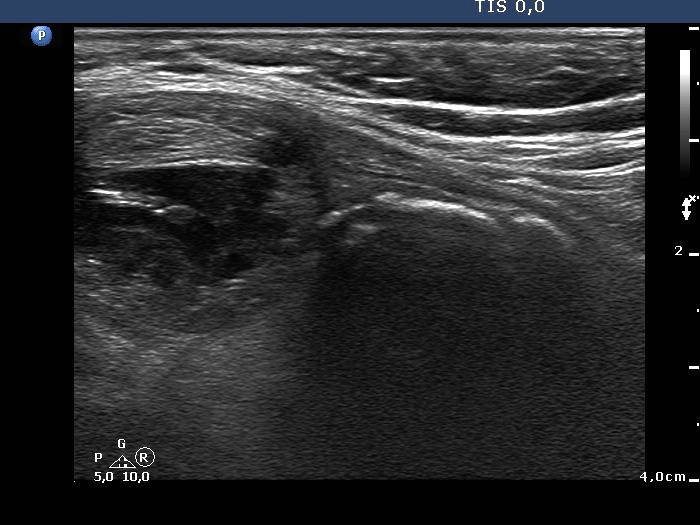
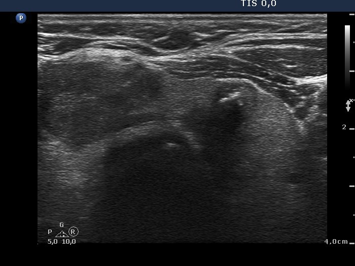
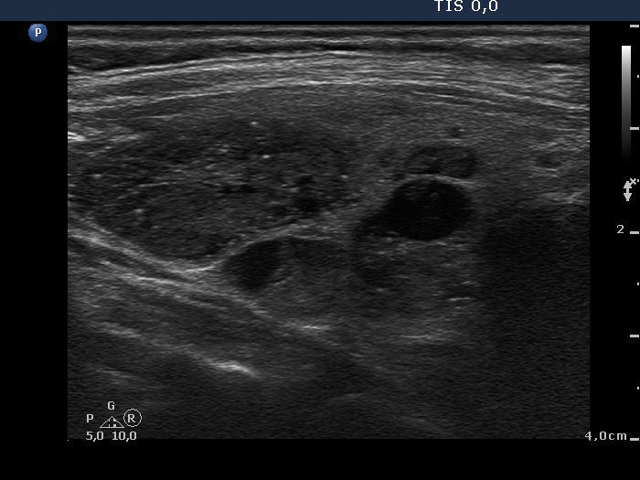
Benign colloid goiter (cytological diagnosis) |
 |
 |
There is a calcification in the dorsal part of the lesion.
|
| |
|
Follicular tumor (cytological diagnosis) - case 1689
|
First examination |
 |
 |
The acoustic shadow begins at the lateral (left image, horizontal view) and lower part (right image longitudinal view) of the nodule. The primary focus can be seen only in the former image. Note hyperechogenic lines and granules at the ventral wall of the lesion.
|
18 months later
|
 |
 |
The calcification present on the original images remained unchanged. The hyperechogenic curved line at the ventral part of the lobe has increased and in contrast with the first examination it seems a rim calcification at the follow-up examination.
|
| |
|
Benign hyperplastic nodule (histological diagnosis) - case 186 |
 |
 |
The lesion presents an incomplete rim calcification.
|
| |
|
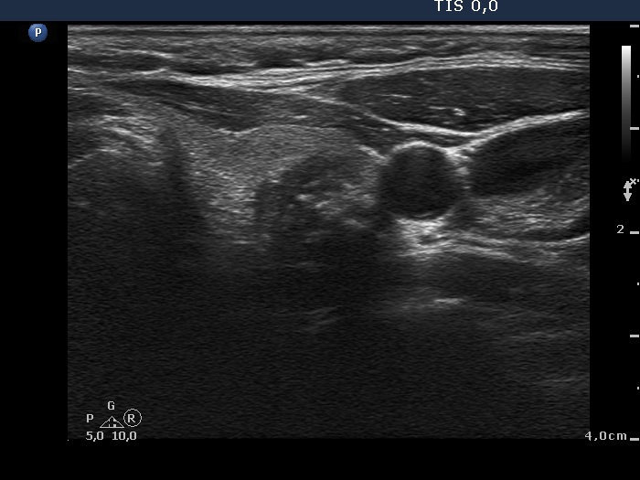
Widely invasive follicular carcinoma - case 20
|
 |
 |
The calcification is within the nodule.
|
| |
|
Papillary carcinoma |
 |
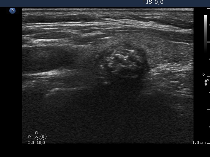 |
The hypoechogenic nodule in the lateral part of the lobe presents coarse calcification and punctate echogenic foci (considering the final diagnosis microcalcifications), too. The proof for the presence of the former is the acoustic shadow. The right image shows pale granules and lines corresponding to normal connective tissue.
|
| |
|
Benign hyperplastic nodule - case 627 |
 |
 |
There is a rim calcification in the ventral part of the nodule.
|
| |
|
Benign hyperplastic nodule - case 653 |
 |
 |
An incomplete eggshell calcification or interrupted peripheral calcification is presented.
|
| |
|
Papillary carcinoma - case 779 |
 |
 |
The tumor has an interrupted peripheral calcification and a few punctate echogenic foci, as well. The hyperechogenic large complex structure in the left, horizontal view is difficult to judge. It might be ragged tissue containing punctate echogenic foci.
|
| |
|
Papillary carcinoma - case 979 |
 |
 |
The tumor presents punctate echogenic foci (microcalcifications) and coarse calcifications, as well.
|
| |
|
|
 |
 |
The presence of coarse calcifications is evident on acoustic shadowing.
|
| |
|
|
 |
 |
The acoustic shadow is the proof for coarse calcification. Moreover, the bright granules in the ventral part of the lesion are punctate echogenic foci.
|
| |
|
Follicular proliferation (cytological diagnosis) - case cons022 |
 |
 |
The lesion has a rim calcification.
|
| |
|
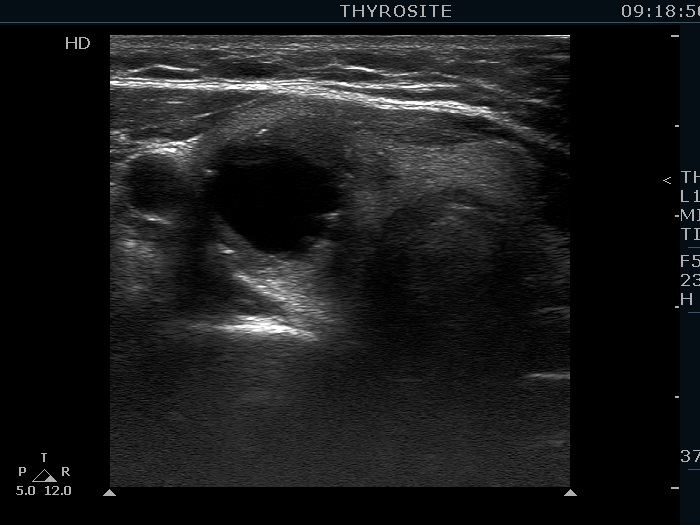
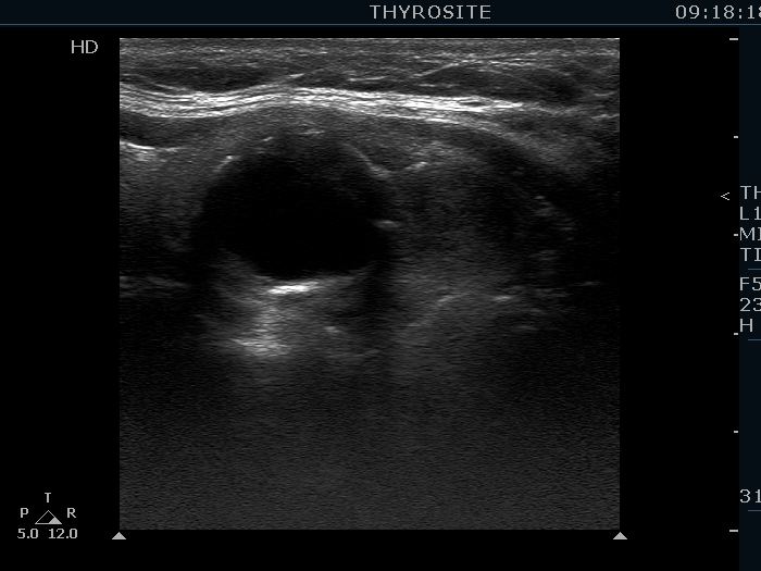
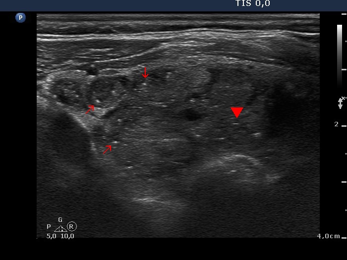
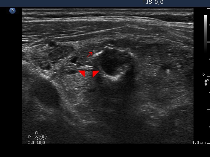
Benign hyperplastic nodule (histological diagnosis) - case cons037 |
Upper part of the right lobe |
 |
 |
 |
 |
The bright hyperechogenic granules (arrows) seem to be at first sight punctate echogenic foci. However, the presence of a few hyperechogenic lines (arrowheads) challenges this view: these figures might be presentations of a connective tissue.
|
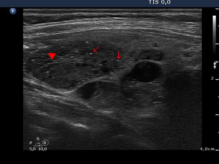
Lower part of the right lobe
|
 |
 |
 |
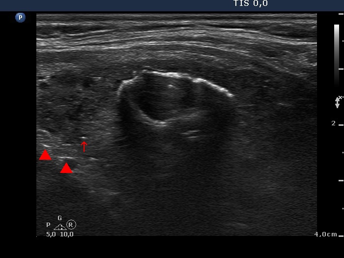 |
This nodule has multiple coarse calcifications and proliferation of a connective tissue (arrowheads and arrows).
|
| |
|
Benign hyperplastic nodule (histological diagnosis) - case cons039
|
 |
 |
Two foci of coarse calcification are presented, one in the left horizontal and another one in the right longitudinal scan.
|
| |
|
Follicular adenoma (histological diagnosis) - case 1519 |
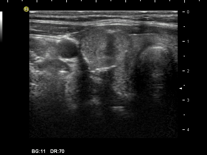 |
 |
The acoustic shadow points to coarse calcifications. On the other hand, the ventral thick hyperechogenic curves only partly correspond to coarse calcification because of the lack of acoustic shadow alongside the figure.
|
| |
|
| |
|
|
