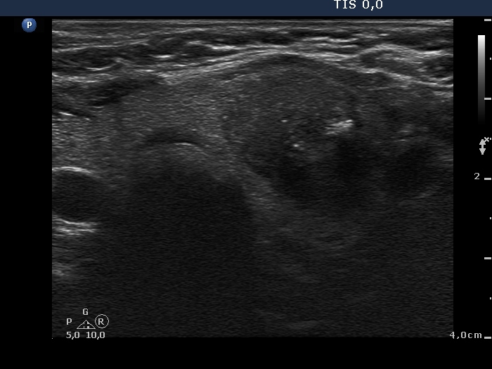
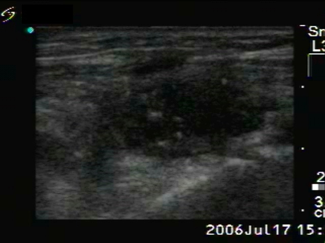
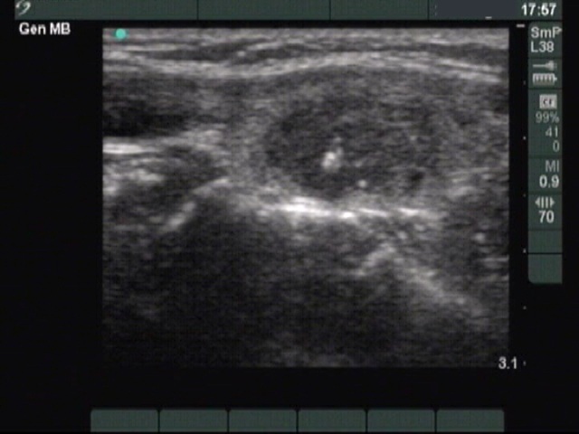
Medullary carcinoma (histological diagnosis) - case 638
|
 |
 |
There is an irregular minimally hypoechogenic-echonormal small patch in the central part of the hypoechogenic nodule. This figure has one bright granule in the left image.
|
| |
|
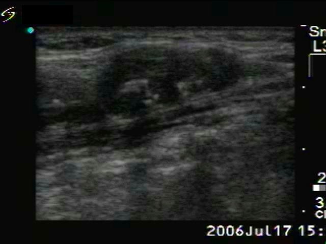
Medullary carcinoma (histological diagnosis)
|
 |
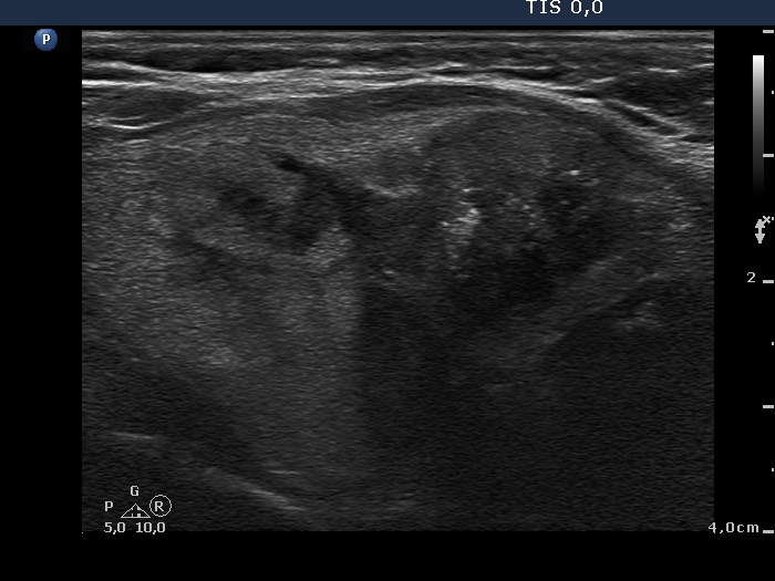 |
The tumor has hyperechogenic granules and a larger echonormal irregularly shaped patch which corresponds to amyloid deposit. Note the dorsal acoustic shadowing.
|
| |
|
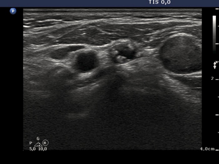
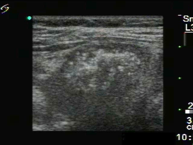
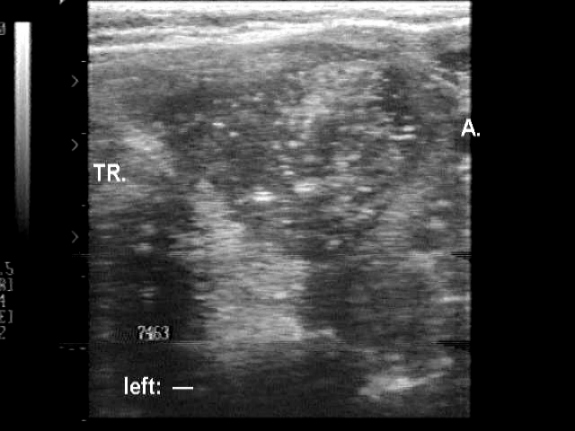
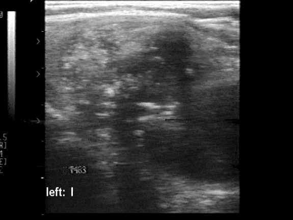
Medullary carcinoma (histological diagnosis) - case 529
|
 |
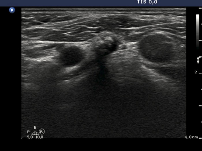 |
A metastatic lymph node is presented in this case. The node has at least four hyperechogenic patches on the left in the horizontal scan. These have an echonormal background and display small hyperechogenic granules. There is acoustic shadow in the right in the longitudinal view.
|
| |
|
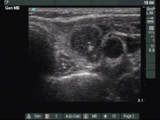
Medullary carcinoma (histological diagnosis) - case 617
|
Right thyroid lobe |
A metastatic lymph node in the right side of the neck |
 |
 |
There are similar hyperechogenic figures in the primary and metastatic focus. These are multiple, have an irregular shape and vary greatly in size.
|
| |
|
Medullary carcinoma (histological diagnosis) - case 1774
|
 |
 |
This tumor contains numerous amyloid deposits. The images were gained with an older equipment, therefore the bright granules within the echonormal background of the patches are larger because of the low quality resolution.
|
| |
|
|
Medullary carcinoma (histological diagnosis) - case 185
|
 |
 |
 |
 |
The papillary carcinoma has punctate echogenic foci (microcalcifications) within an echonormal background which is itself a thyroid tissue.
|
The bright granules are located within an echonormal background. These together are the amyloid deposits.
|
| |
|
Medullary carcinoma (histological diagnosis) - case 174
|
 |
 |
This tumor has numerous bright punctate echogenic foci and large irregularly shaped echonormal patches, the latter are amyloid deposits.
|
| |
| |
| |
| |
| |
|
