
|
|
Benign nodular hyperplasia - Case 9.
|
|


Clinical presentation: A 65-year-old woman was referred for evaluation of a goiter. A nodule in the left lobe has been known for more than 20 years. The patient noticed a new lump in the right side of the neck which caused dysphagia.
Palpation: an elastic nodule in the right and a hard nodule in the left lobe.
Functional state: euthyroidism with TSH-level 2.09 mIU/L.
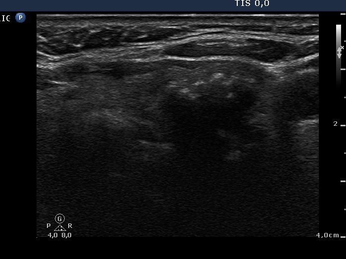
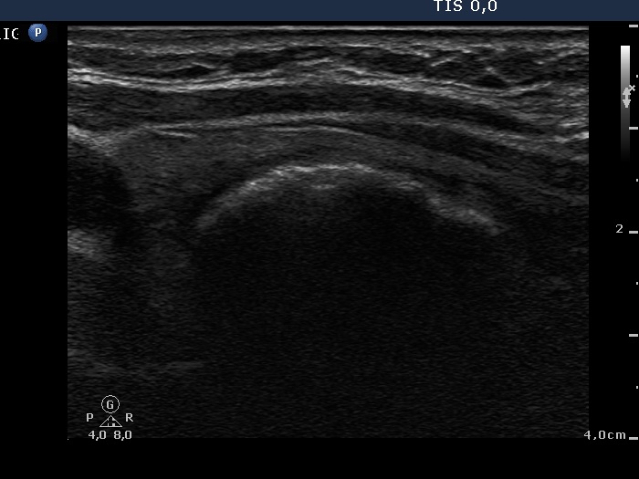
Ultrasonography: There was a large cystic nodule in the right while a large nodule with eggshell calcification in the left lobe.
Cytology from the nodule in the right lobe was repeatedly not diagnostic. We aspirated 32 mL brown fluid. The nodule refilled next day after the evacuation. The FNA of the nodule in the left lobe (see cytological images) resulted in microfollicular proliferation.
Combined sonographic-cytological diagnosis: benign lesions with great probability.
Surgery was advised and a total thyroidectomy was performed.
Histopathology: benign, hyperplastic nodular goiter.
Comment: This is a very good example for a need for a new diagnosing-reporting system. Follicular proliferation causes the major problem in thyroid cytology. Not only follicular adenomas and follicular carcinomas belong to this group, but also hyperplastic nodules and Hashimoto's thyroiditis can display this pattern. In this case, the cytological picture may correspond to a follicular tumor but the lack of halo sign and perinodular blood flow are strong arguments against a follicular tumor. If we take the sonographic pattern into account, we can avoid the false diagnosis of a follicular tumor.