|
|||||||||||||||||||||||||||||||||||||||||||||||||||||||||||||||||||||||||||||||||||||||||||||||||||
Selected topics - intranodular hyperechogenic figures - Table 1. Coexistent echogenic lines and granules |
|||||||||||||||||||||||||||||||||||||||||||||||||||||||||||||||||||||||||||||||||||||||||||||||||||
|
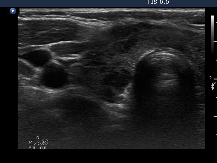
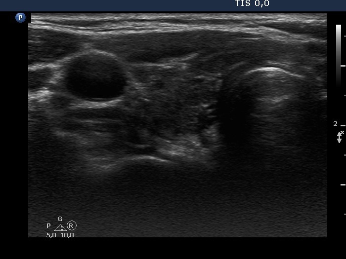
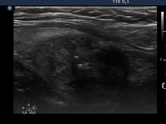
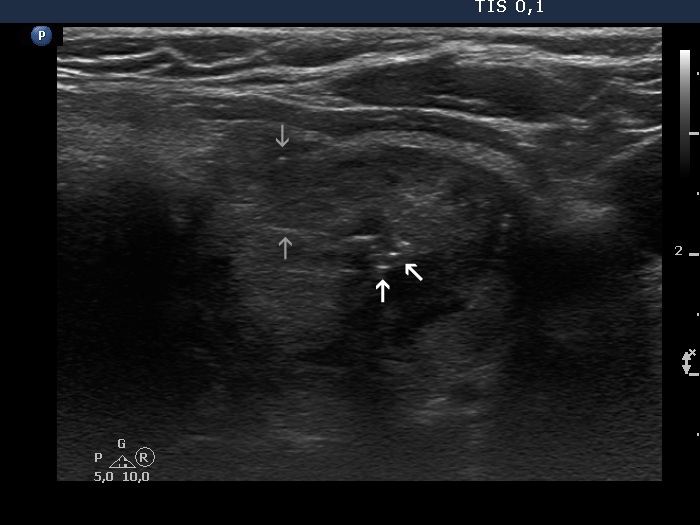
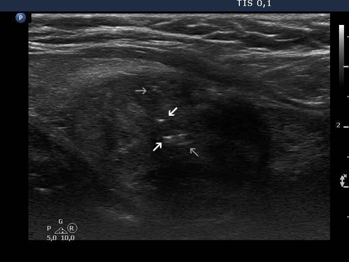
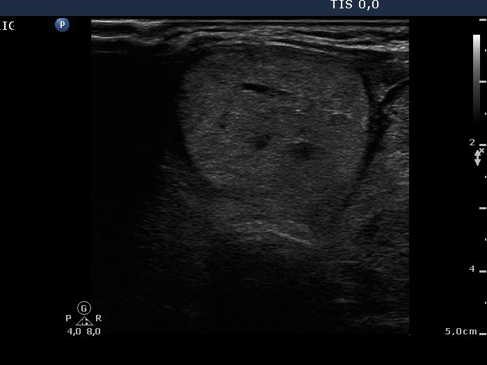
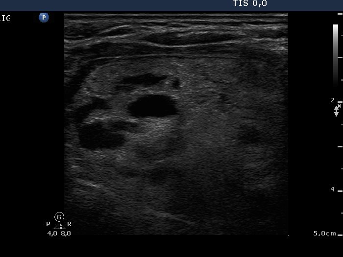
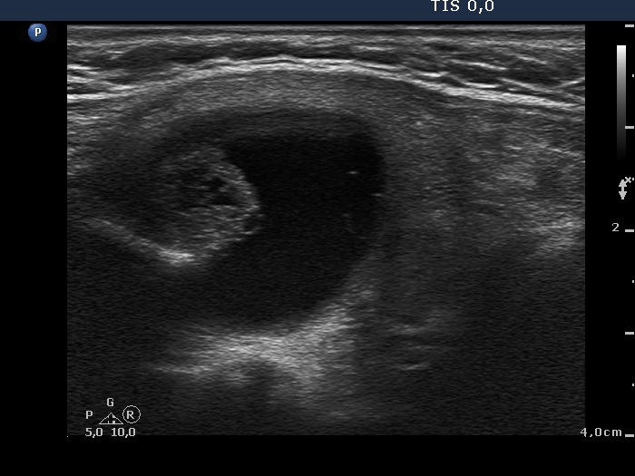
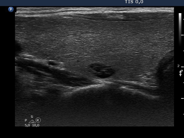
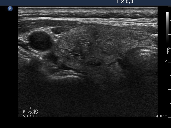
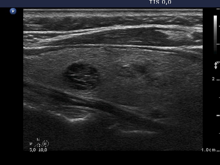
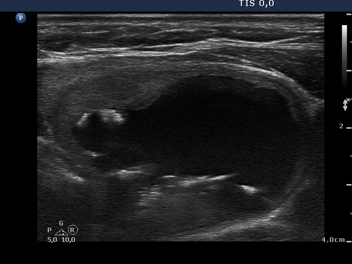
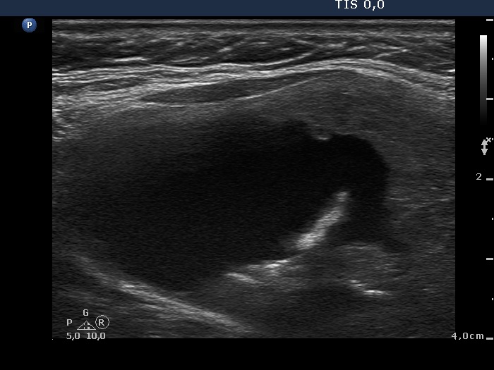
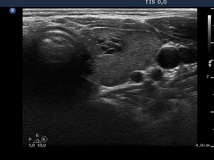
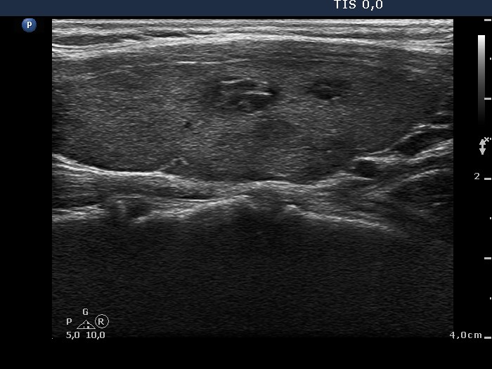
Powdery tiny and usually pale granules and short lines are the most frequent subtype of intranodular figures. These are normal findings and represent normal pattern of a connective tissue. Occasionally they appear in the form of a bit brighter granules and lacking lines. In that case the differentiation from punctate echogenic foci including microcalcification is not possible. Occasionally large thick, amorphous strings are found which correspond to thick connective tissue. In the event of proliferation of connective tissue (or fibrosis) more bright granules and longer lines occur simultaneously. The presentation is very similar to back wall cystic figures. Because the wall of cystic areas is composed of connective tissue, it is not surprising that the differentiation between these two subgroups is not always possible. In contrast with the former issue which shares only a theoretical problem, the differentiation from punctate echogenic focus (microcalcification) is a real concern. If we overlook the hyperechogenic lines which are always present in fibrosis, bright and relatively larger granules are easily misinterpreted as punctate echogenic foci (microcalcifications). |
|||||||||||||||||||||||||||||||||||||||||||||||||||||||||||||||||||||||||||||||||||||||||||||||||||
|
|||||||||||||||||||||||||||||||||||||||||||||||||||||||||||||||||||||||||||||||||||||||||||||||||||