
Benign nodular hyperplasia - Case 59. (ultrasonographic picture 5)
|
|

|
|
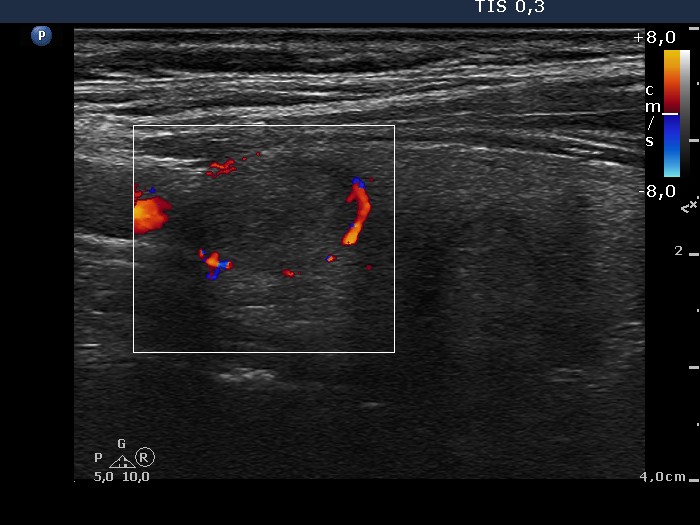
Upper part of the right lobe, longitudinal scan, color Doppler mode. The nodule in the upper part presents perinodular blood flow.