
Benign nodular hyperplasia - Case 59. (ultrasonographic picture 6)
|
|

|
|
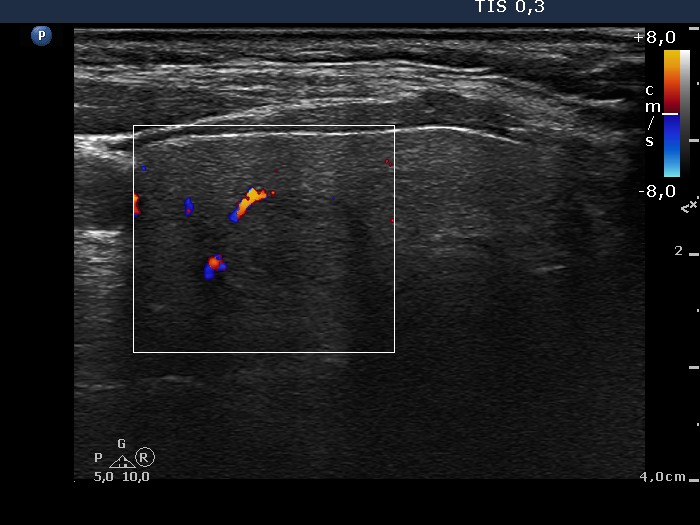
Middle-lower part of the right lobe, horizontal view, color Doppler mode. This lesion also displays signs of perinodular blood flow.