
|
|
Benign nodular hyperplasia - Case 59. |
|


Clinical presentation: a 41-year-old woman was referred for follow-up evaluation of a nodular goiter. Previous cytology resulted in benign colloid goiter. She had no complaints.
Palpation: multiple not firm nodules in the right lobe.
Functional state: euthyroidism (TSH 0.57 mIU/L).
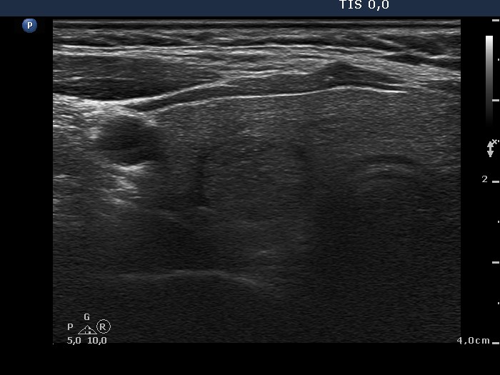
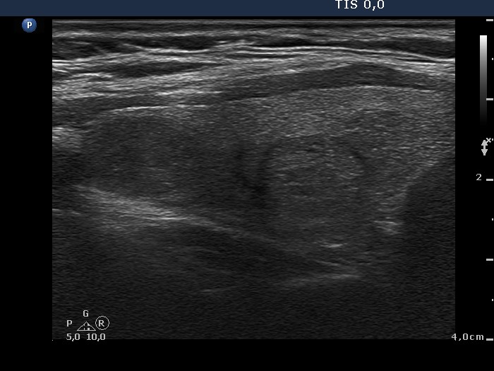
Ultrasound. The thyroid was echonormal. There were two lesions in the right lobe, the upper one was minimally hypoechogenic and had blurred borders while the nodule in the middle part of the lobe was inhomogeneous and displayed halo. Both nodules presented perinodular blood flow. The upper lesion was not noticed in the previous examination 4 years ago.
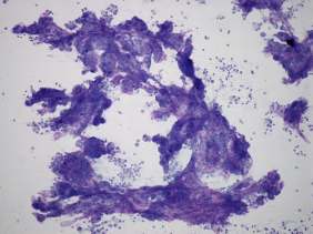
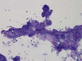
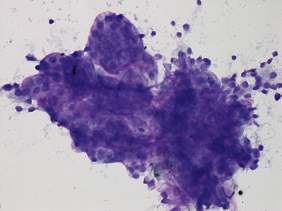
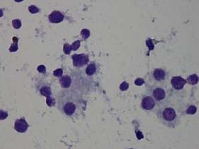
Aspiration cytology was performed from the nodule in the upper part of the right lobe. Our cytological report was suspicion of papillary carcinoma with around 10% risk of malignancy.
Histopathology disclosed benign hyperplastic nodules with focal papillary hyperplasia.
Comments.
-
Cell balls are characteristic signs of a hyperplastic nodules. These are commonly found isolated. In this case the cell balls formed papillary-like clusters which is an unusual finding. There were a few atypical cells on the smear; this is a very common finding in benign thyroid lesions. All in all, the cytological pattern itself corresponds to a benign hyperplastic lesion.
-
The lesion in question was not present four years earlier and had blurred borders. Both of these circumstances increased the likelihood of malignancy.
-
Taking all these into account, I gave a wrong diagnosis.