
|
|
Chronic lymphocytic thyroiditis - Case 48.
|

|




Clinical data: a 53-year-old woman was referred for an evaluation of a 'nodular goiter'.
Palpation: both lobes were firm, nodular.
Functional state: euthyroidism (TSH 3.30 mIU/L).
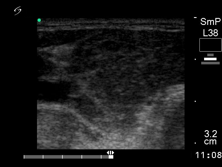
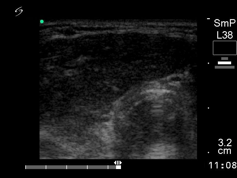
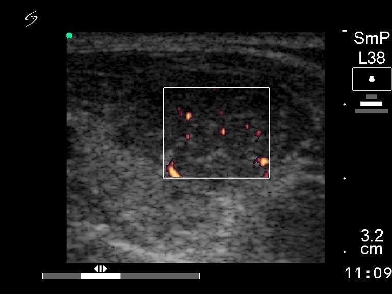
Ultrasonography: hypoechogenic thyroid with several circumscribed lesions corresponding to Hashimoto's thyroiditis.
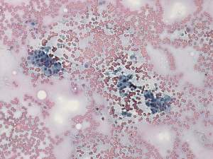
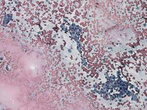
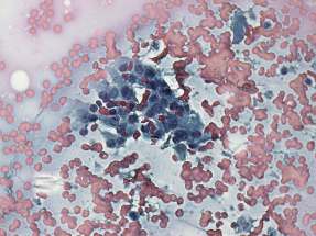
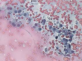
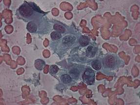
Cytological picture: there is no colloid in the background. Thyrocytes exhibiting oxyphilic metaplasia are in small non-specific clusters. Only a few lymphocytes are present in the background. A cell with two inclusions can be found.
Combined clinical-cytological diagnosis: Hashimoto's thyroiditis.
We performed anti-TPO which yielded 709 IU/mL.
Suggestion: follow-up examination. Hypothyroidism developed two years later and the thyroid became atrophic.


