
|
|
Follicular adenoma - Case 29.
|
|


Clinical data: a 34-year-old man was referred for an evaluation of a thyroid nodule that had been discovered 2 years earlier.
Palpation: a nodule in question in the right lobe.
Functional state: euthyroidism with TSH-level of 1.88 mIU/L.
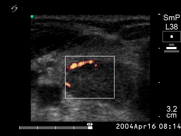
Ultrasonography: an echonormal right lobe with several small hypoechogenic lesions and a hypoechogenic nodule in the dorsal part. The nodule did not exhibit a halo sign but on Doppler mode perinodular blood flow was detected.
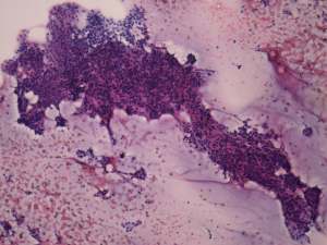
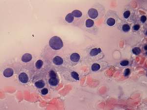
Cytological picture: there is no colloid in the background. Thyrocytes forming microfollicles and small clusters. No significant atypia is present.
Cytological diagnosis: follicular tumor with less than the average risk for malignancy.
Histopathological diagnosis: dominantly normofollicular adenoma. Chronic lymphocytic thyroiditis in the extranodular part of the lobe.
Comment: although the nodule was non palpable, it caused an unevenness of the ventral surface of the lobe. This is a very good example why is US-guided aspiration superior to palpation guided. In this patient a palpation guided aspiration would be performed with great probability from the non-nodular part of the lobe.