
|
|
Papillary carcinoma - Case 35.
|
|


Clinical data: a 27-year-old woman came to a regular follow-up examination. A small lesion was detected 10 years earlier with a maximal diameter of 8 mm. We met the patient first 5 years later when a nodule with the dimensions of 13x10x18 mm was diagnosed. Aspiration cytology resulted in follicular proliferation. We advised yearly follow-up instead of immediate surgery because no atypia was present of the smear. The patient came to the next follow-up examination only 5 years later. She told that her nodule has increased in size in the last 6 months.
Palpation: a nodule was palpable in the right lobe.
Functional state: euthyroidism (TSH-level 2.14 mIU/L).
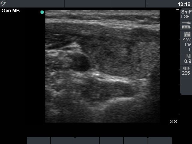
Ultrasonography: revealed a moderately hypoechogenic nodule with a halo sign in the right lobe. The dimensions of the right nodule increased from 13x10x18 mm to 21x16x30 mm over 5 years.
Cytology.
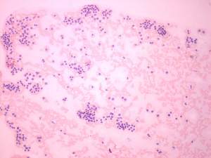
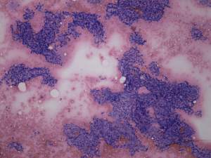
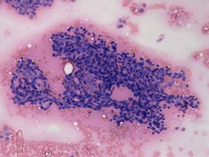
First examination, 5 years earlier (first row of cytological images)
Diagnosis: follicular tumor.
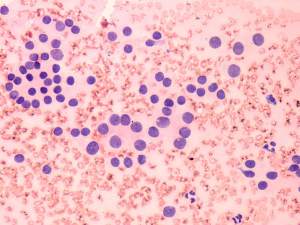
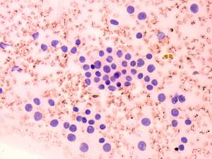
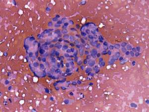
Present examination (second row of cytological images)
Diagnosis: papillary carcinoma.
Surgery was advised.
Histopathology: encapsulated papillary carcinoma dominantly follicular variant.
Comments:
1. This is one of our few cases where we did not advised surgery in the case of a benign appearing follicular proliferation and the final histopathology disclosed carcinoma. Although the appropriate treatment was performed 5 years later, the patient did not suffer any harm. There were no metastatic foci and the serum-thyroglobulin level is undetectable after thyroidectomy and radioiodine therapy.
2. By reviewing the original smears we could not find any cytological signs suggesting papillary cancer. This is an extremely unusual situation.