
|
|
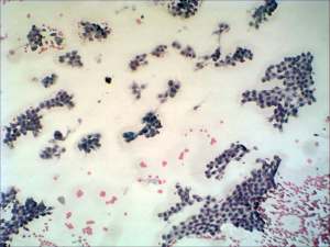
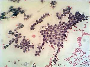
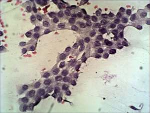
Papillary carcinoma - Case 62.
|
|


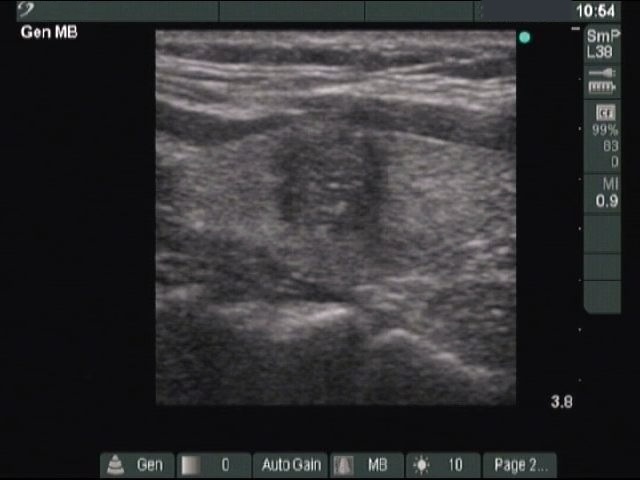

Clinical data: a 37-year-old woman with a thyroid nodule discovered on carotid Doppler ultrasonography.
Palpation: no abnormality on palpation.
Functional state: euthyroidism (TSH 0.91 mIU/L).
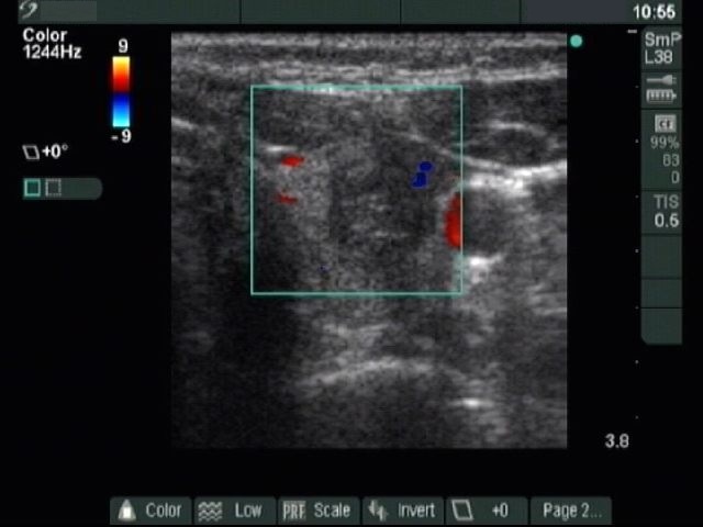
Ultrasonography: the thyroid was echonormal. There was a hypoechogenic nodule in the left lobe which showed microcalcifications and had irregular borders. Neither intra- nor perinodular blood flow was present.
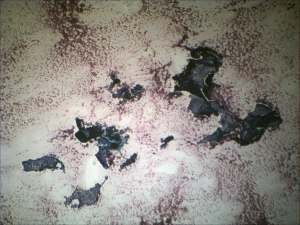
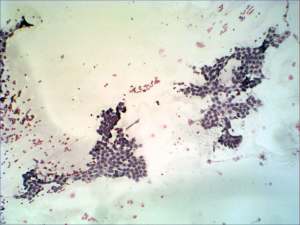
Cytological picture: there was no colloid in the background. Thyrocytes in monolayered sheets. Occasionally nuclear crowding can be observed. Thyrocytes exhibit oxyphilic metaplasia. Many cells contain groove, nuclear inclusion are present occasionally.
Cytological diagnosis: suspicion of papillary cancer.
Histopathology: papillary cancer.