
|
|
Papillary carcinoma - Case 61.
|
|


Clinical data: a 28-year-old woman requested a second opinion. She was previously evaluated because of clinical suspicion of hyperthyroidism (irritability, weight loss, neck problems), but hormonal evaluation excluded hyperthyroidism. No ultrasonography was performed.
Palpation: the right lobe was moderately firm and the presence of a nodule was questionable.
Functional state: euthyroidism (TSH 1.07 mIU/L).
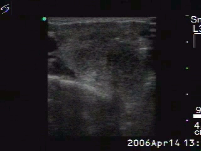
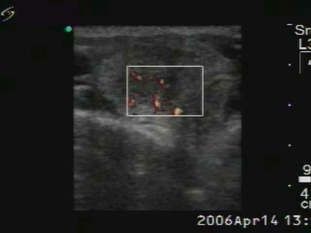
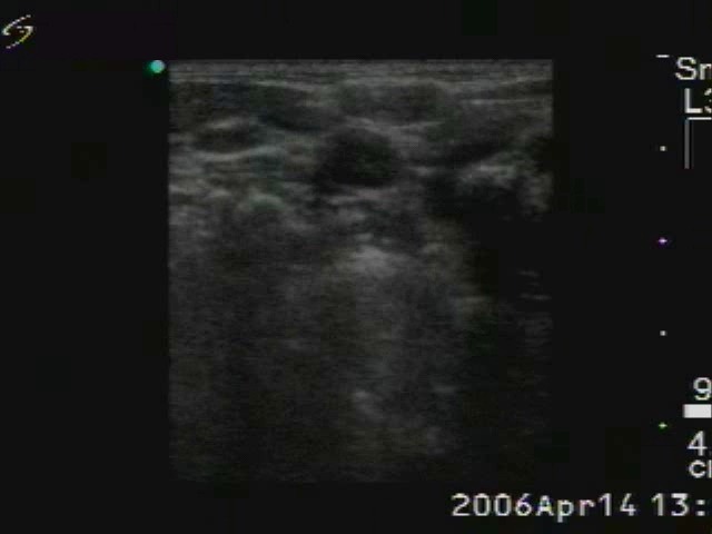
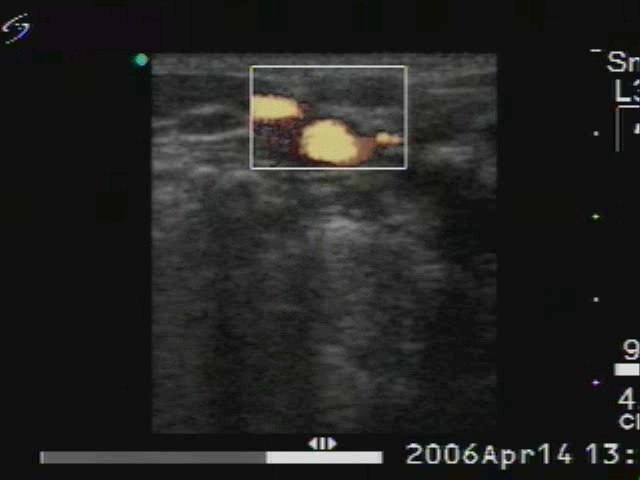
Ultrasonography: the thyroid was echonormal. There was a relatively large hypoechogenic area in the right lobe with blurred borders. The vascularization within the nodule was increased.
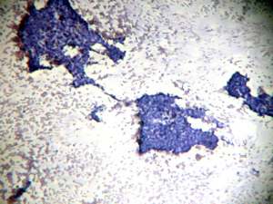
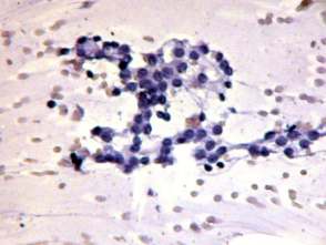
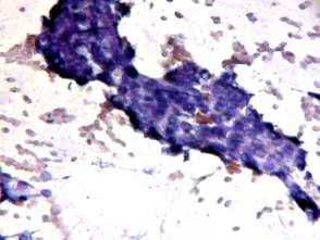
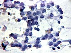
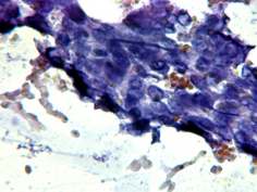
Cytological picture: there was no colloid in the background. Cells are arranged in non-specific groups, loose papillary structures with nuclear crowding and overlapping. Because of the degenerative changes, the nuclear details were not unequivocal but we have found intranuclear structures resembling inclusions and grooves.
Cytological diagnosis: suspicion of papillary cancer.
Histopathology: papillary cancer.