
Benign nodular hyperplasia - Case 27. (ultrasonographic picture 4)
|
|

|
|
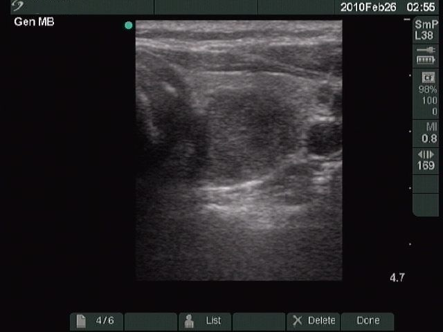
Left lobe, horizontal scan. There is a hypoechogenic, inhomogeneous nodule on the dorsal surface of the lobe. The white punctures within the nodule do not correspond to microcalcification but to connective tissue. The ventro-lateral border of the nodule is a little bit blurred.