
Benign nodular hyperplasia - Case 27. (ultrasonographic picture 6)
|
|

|
|
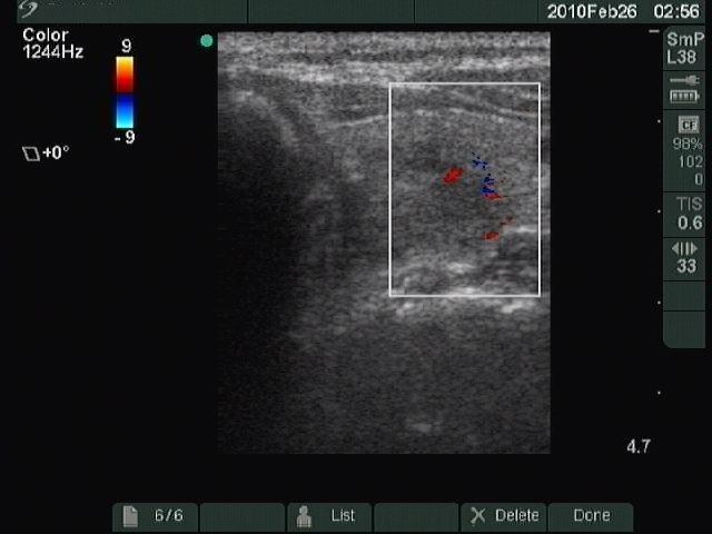
Left lobe, horizontal scan, color Doppler method. The left larger red patch corresponds to an intranodular vessel, while the other powder-like blue and red points are artifacts.