
|
|
Papillary carcinoma - Case 59.
|
|


Clinical data: a 45-year-old man requested an evaluation because both parents had hypothyroidism. He had no complaints.
Palpation: no abnormality.
Functional state: euthyroidism (TSH 1.82 mIU/L).
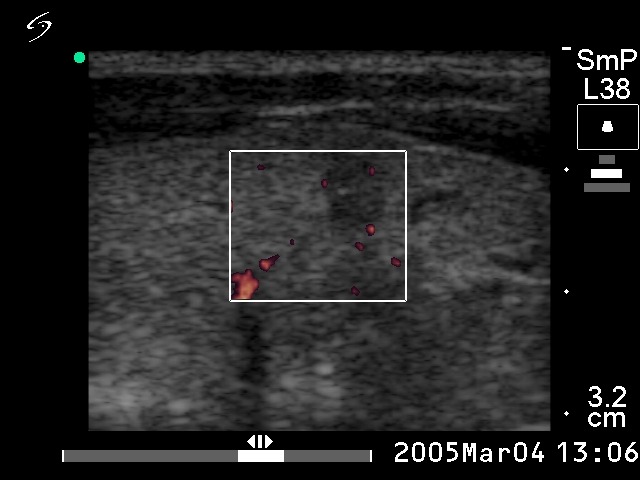
Ultrasonography: the thyroid was echonormal. There were two small hypoechogenic lesions close to each other in the medial part of the right lobe. One of them with a maximal diameter of 4 mm contained microcalcification. The vascular pattern in such small lesions has no relevance.
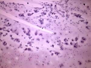
Cytological picture: the presence of colloid is equivocal. Cells are arranged in non-specific groups and occur dissociated. The follicular cells have abundant cytoplasm and many of them display inclusion. Multinucleated cells and lymphocytes were also present. The background was dirty. A psammoma body was also found. Cytological diagnosis: papillary cancer.
Histopathology: papillary microcancer with a maximal diameter of 4 mm.