
|
|
Papillary carcinoma - Case 58.
|
|


Clinical data: a 65-year-old woman was referred for an evaluation of a rapidly growing mass in the thyroid discovered 2 months earlier.
Palpation: a hard nodule in the isthmus.
Functional state: euthyroidism (TSH-level 0.76 mIU/L).
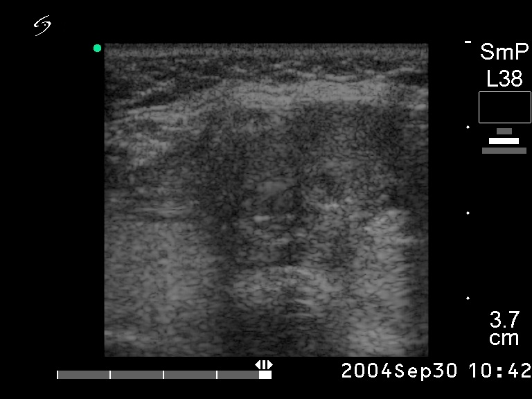
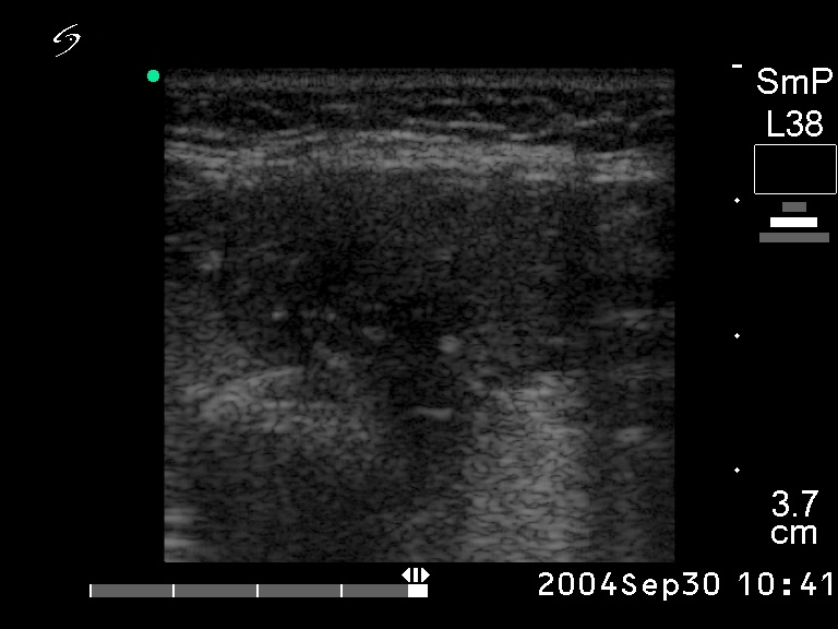
Ultrasonography: the basic echo structure of the thyroid was normal. There were 3 lesions in the right, while 1 lesion in the left lobe. The hypoechogenic one in the upper pole of the right lobe was aspirated.
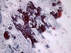
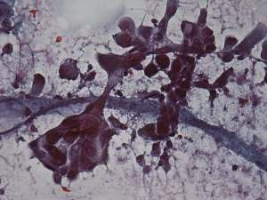
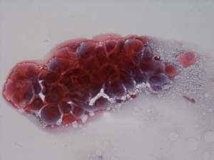
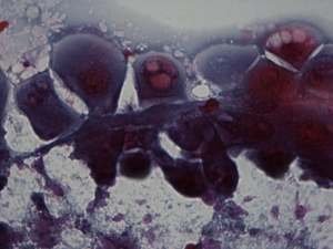
Cytological picture: no colloid in the background. Irregular clusters with nuclear crowding and overlapping, several typical papillary fronds. The thyrocytes exhibit enlargement, prominent nucleoli, oxyphilic metaplasia and occasionally squamous metaplasia. No grooves or inclusions can be identified.
Cytological diagnosis suspicion of papillary carcinoma.
Histopathology: papillary carcinoma.